Micro-fluidic device and method for detecting circulating tumor cell on basis of dimension

A microfluidic device and tumor cell technology, which is applied in the field of microfluidic devices for size-based detection of circulating tumor cells, can solve the problems of low detection sensitivity, complicated detection process, and long detection time of circulating tumor cells, so as to improve sensitivity, The effect of low precision requirement and low production cost

Active Publication Date: 2016-11-09
SHANGHAI INST OF MICROSYSTEM & INFORMATION TECH CHINESE ACAD OF SCI
View PDF9 Cites 44 Cited by
  • Summary
  • Abstract
  • Description
  • Claims
  • Application Information

AI Technical Summary

Problems solved by technology

[0008] The purpose of the present invention is to provide a microfluidic device and method for detecting circulating tumor cells based on size, so as to solve the defects of low detection sensitivity, complicated detection process, high cost and long time-consuming detection of circulating tumor cells in the prior art

Method used


Image

  • Micro-fluidic device and method for detecting circulating tumor cell on basis of dimension
  • Micro-fluidic device and method for detecting circulating tumor cell on basis of dimension
  • Micro-fluidic device and method for detecting circulating tumor cell on basis of dimension

Examples


Embodiment 1

[0042] Example 1 Preparation of microfluidic chip

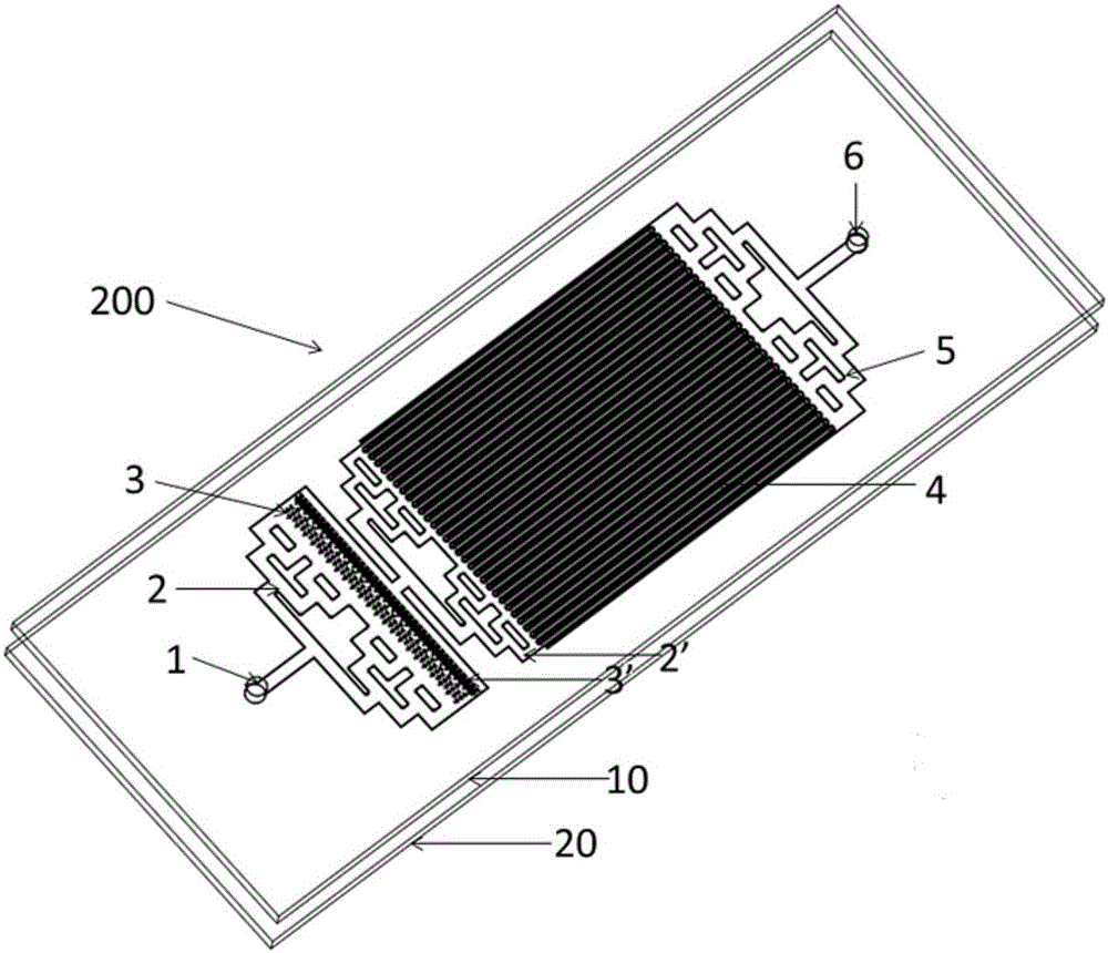
[0043] 1.1 According to the above-mentioned chip structure designed in the present invention, utilize AutoCAD software to draw out required figure, make film mask plate; With four-inch monocrystalline silicon chip as substrate, carry out thermal oxidation to form an oxide layer of one deck 2 μm, then carry out Photolithography, reactive ion etching, adhesive stripping and cleaning, photolithography, deep reactive ion etching, etching out the height of the filter channel 43 . Concentrated H 2 SO 4 Wash with hydrogen peroxide, remove the photoresist, perform deep reactive ion etching according to the silicon dioxide pattern again, etch out the height of other channels of the chip, and finally obtain a silicon wafer mold with a microstructure.

[0044] 1.2 Place the silicon wafer mold and the open centrifuge tube containing 10 μL of fluorosilane in a vacuum drying oven, vacuum to negative pressure to vaporize the fluorosilane,...

Embodiment 2

[0046] Example 2 Using the microfluidic chip prepared in Example 1 to detect circulating tumor cells

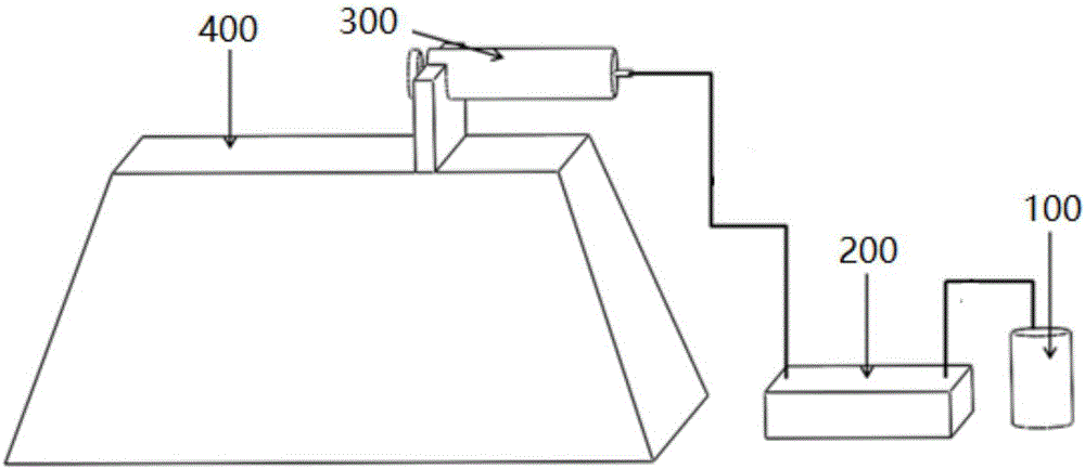
[0047] First, centrifuge 2 mL of blood sample, absorb the upper layer of plasma, and then lyse the remaining red blood cells twice, then resuspend the remaining cells with 500 μL of 4% paraformaldehyde solution, fix the cells, and finally store them in a 4°C refrigerator for later use. For detection, first draw 250 μL of the treated cell suspension and dilute it to 1 mL for later use. The flow rate of the sampling pump 400 is set to 15mL / L, and the diameter is set to 19mm. One end of the waste liquid collection syringe 300 is connected to the sample outlet 6 of the microfluidic chip 200. The sample inlet 1 of the fluidic chip 200 draws the sample into the microfluidic chip 200; then washes the chip with 200 μL of PBS (containing 0.05% Tween) washing solution, and the smaller red blood cells and white blood cells are washed out of the microfluidic chip. The chip 200 enters th...

Embodiment 3

[0049] Using the device provided in Example 1 and the detection method provided in Example 2, the flow rate of the sampling pump was set to 10mL / h, 15mL / h, and 17mL / h respectively, and the lung cancer cell line H446 suspension of known concentration was tested. Calculate the capture efficiency of H446 cells at different flow rates, see the experimental results Figure 5 , as can be seen from the figure, the capture efficiency of the microfluidic chip is the highest at a flow rate of 15mL / h, as high as 94%.


PUM

PropertyMeasurementUnit
diameteraaaaaaaaaa

Abstract

The invention provides a micro-fluidic device and a method for detecting circulating tumor cells on the basis of dimensions. The micro-fluidic device comprises a solution storage chamber, a micro-fluidic chip, a waste liquid collecting syringe and a power system which are connected in sequence, wherein the micro-fluidic chip is made of a glass substrate layer and a PDMS (Polydimethylsiloxane) chip layer in a laminated manner; the PDMS chip layer comprises an injection port, a block mass filtering area, a target sell screening area and a discharge port which are communicated in sequence; the block mass filtering area is formed by a column array; the target cell screening area comprises a plurality of main pipelines and side pipelines which are extended in a parallel manner at intervals and are communicated through filtering channels; the front ends of the main pipelines are opened; the filtering channels are formed in the rear ends of the main pipelines; the front ends of the side pipelines are sealed; the rear ends of the side pipelines are opened; the main pipelines and the side pipelines have a first height greater than the dimensions of the circulating tumor cells; the filtering channels have a second height smaller than the dimensions of the circulating tumor cells. The micro-fluidic device and the method for detecting the circulating tumor cells on the basis of the dimensions are good in sensitivity, simple to operate, low in cost, large in flux and short in time.

Description

technical field [0001] The invention relates to the field of biological detection, in particular to a microfluidic device and method for size-based detection of circulating tumor cells. Background technique [0002] Cancer is still one of the diseases with the highest mortality rate in the world. In 2008, 7.6 million people died from cancer (13% of all deaths), and by 2030 it is likely to exceed 13 million. Early diagnosis and treatment of primary cancer and effective treatment of metastatic cancer are the key to fighting cancer. [0003] Circulating tumor cells (CTCs) are a type of tumor cells that enter the blood circulation system from the primary tumor. Such cells can transfer through the blood circulation system and reach other organs to achieve tumor metastasis, so CTCs are closely related to the occurrence, development and metastasis of tumors. In view of the important role of CTCs in tumor metastasis, we can monitor the effect of tumor treatment and recurrence in ...

Claims


Application Information

Patent Timeline
Patent Type & Authority Applications(China)
IPC IPC(8): B01L3/00G01N1/30G01N33/533
CPCB01L3/502761B01L2300/0809G01N1/30G01N33/533
Inventor 贾春平郜晚蕾李刚景奉香金庆辉赵建龙
Owner SHANGHAI INST OF MICROSYSTEM & INFORMATION TECH CHINESE ACAD OF SCI
Why Eureka
Try Eureka