Time-resolved fluorescence immunochromatographic reagent for rapid quantitative simultaneous detection of cTnI, CKMB, Myo and preparation method

A time-resolved fluorescence and immunochromatography technology, applied in the field of clinical medical diagnosis, can solve the problems of background signal interference, inability to meet the sensitivity, multi-sample size, etc., to improve the detection precision and accuracy, reduce the background signal value, eliminate the The effect of sample interference

Inactive Publication Date: 2016-11-23
SHANGHAI UPPER BIO TECH PHARMA
View PDF10 Cites 22 Cited by
  • Summary
  • Abstract
  • Description
  • Claims
  • Application Information

AI Technical Summary

Problems solved by technology

Colloidal gold immunochromatography for qualitative or quantitative detection has low sensitivity and narrow linear range, which cannot meet the requirements of quantitative and accurate detection; microfluidic method can quantitatively, accurately, and simultaneously detect the three items of myocardium, but often requires a large sample size , and the cost of reagents is high; most fluorescent immunochromatography methods use fluorescein or other substances as light sources. Although they have a high linear range, they cannot guarantee low-end sensitivity due to the interferen

Method used


Image

  • Time-resolved fluorescence immunochromatographic reagent for rapid quantitative simultaneous detection of cTnI, CKMB, Myo and preparation method
  • Time-resolved fluorescence immunochromatographic reagent for rapid quantitative simultaneous detection of cTnI, CKMB, Myo and preparation method
  • Time-resolved fluorescence immunochromatographic reagent for rapid quantitative simultaneous detection of cTnI, CKMB, Myo and preparation method

Examples


Embodiment 1

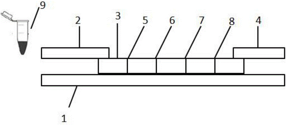
[0061] Preparation of cTnI, CKMB, Myo time-resolved fluorescent immunochromatographic test strips:

[0062] (1) Preparation of detection line (T1, T2, T3) solution: dilute cTnI monoclonal antibody and polyclonal antibody, CKMB polyclonal antibody, and Myo monoclonal antibody to 1-2 mg with 10-fold, 50 mM citrate solution. / ml;

[0063] (2) Preparation of the quality control line (C) solution: dilute the rabbit IgG antibody to 1-2 mg / ml with 10-fold, 50 mM citrate solution;

[0064] On the bottom plate (1) with adhesive backing, adopt the overlapping method, first paste the nitrocellulose membrane (3), and then paste the Fusion5 membrane (2) and the absorbent paper ( 4). On the nitrocellulose membrane (3) and close to the Fusion5 membrane (2), draw T1 (Myo capture line (5)), T2 (cTnI capture line (6)), T3 (CKMB capture line (7)), respectively, Draw a C (rabbit IgG) line near one end of the absorbent paper (4), and the distances between T1, T2, T3, and C are all 3mm.

[0065...

Embodiment 2

[0068] Detection of cTnI, CKMB, Myo time-resolved fluorescent immunochromatographic test strips

[0069] (1) Preparation of cTnI, CKMB, Myo, goat anti-rabbit time-resolved fluorescent microspheres

[0070] Dissolve time-resolved fluorescent microspheres (particle size about 200nm) in 100mM MES buffer (pH 6.0), add activator (NHS, 20mg / ml; EDC, 20mg / ml) to activate for 15 minutes; wash, centrifuge, Reconstitute the time-resolved fluorescent microspheres with 60mM boric acid buffer (pH 8.5), then add troponin I monoclonal antibody 2 to react for 2 hours (the mass ratio of microspheres to antibodies is 10mg:0.5mg); the reaction is completed Then add blocking agent (BSA, 100mg / ml) to block for 2 hours; after blocking, wash, centrifuge, redissolve with 60mM boric acid buffer (pH 8.5) and blocking agent (BSA, 100mg / ml) again, sonicate, make The microspheres are evenly dispersed in the buffer, and stored at 2-8°C in the dark.

[0071] The preparation process of fluorescent microsph...

Embodiment 3

[0084] Precision testing

[0085] With the test method in embodiment 2, test the mixed standard substance of Myo, cTnI and CKMB respectively (the concentration of Myo is 100 and 400ng / ml, the concentration of cTnI is 1 and 25ng / ml, the concentration of CKMB is 10 and 50ng / ml) , and each standard product was repeatedly detected ten times, and the specific detection values ​​are shown in Table 2 below.

[0086] Table 2 precision test result table

[0087]

[0088] As can be seen from the data in Table 2, using the triple detection test strip of the present invention, the precision of Myo, cTnI, and CKMB is all less than 10%, which fully meets the requirement that the precision of POCT products is less than 15%.


PUM

PropertyMeasurementUnit
Particle sizeaaaaaaaaaa
Sensitivityaaaaaaaaaa

Abstract

The invention relates to the field of clinical diagnosis, in particular to a time-resolved fluorescence immunochromatographic reagent for rapid quantitative simultaneous detection of cTnI, CKMB and Myo, and a preparation method thereof. The reagent comprises a test strip and fluorescent liquid, wherein the test strip comprises a bottom plate, Fusion5, a nitrocellulose membrane and an absorbent pad, and the Fusion5, the nitrocellulose membrane and the absorbent pad are sequentially connected and fixed on the bottom plate in the horizontal direction. Time-resolved fluorescent microspheres are used to improve the fluorescence intensity and decrease background signals, and cTnI, CKMB and Myo indexes in whole blood, serum or plasma can be simultaneously and quantitatively detected, and a sample needs only 10 to 20mul. The test strip has the advantages of convenience, rapidness, simple operation, short detection time, strong specificity, high sensitivity and relatively accurate test results, and is suitable for rapid diagnosis of clinical POCT.

Description

technical field [0001] The invention belongs to the field of clinical medical diagnosis, in particular to a time-resolved fluorescent immunochromatographic reagent for rapid quantitative simultaneous detection of cTnI, CKMB and Myo and a preparation method thereof. Background technique [0002] Troponin is a complex composed of three subunits, troponin T (cTnT), troponin C (cTnC), and troponin I (cTnI). The molecular weight of cTnI is 22.5kD. The N-terminus of the polypeptide has 26 more amino acids than skeletal muscle TnI, and the difference in amino acid sequence is 40%, so the antigenicity is obviously different from that of skeletal muscle. As a new diagnostic marker, cTnI is used in the clinical diagnosis of acute myocardial infarction (AMI), especially for those patients without electrocardiographic diagnosis. When AMI occurs, cTnI will be released from the injured cardiomyocytes into the blood within 4 to 8 hours, and the concentration exceeds the normal range. Usu...

Claims


Application Information

Patent Timeline
IPC IPC(8): G01N33/577G01N21/64G01N33/68
CPCG01N33/577G01N21/6408G01N33/6887
Inventor 朱传增石晓强朱奇朗安永君张蕾
Owner SHANGHAI UPPER BIO TECH PHARMA
Why Eureka
Try Eureka