drug-coated stent graft

A covered stent and drug application technology, applied in the field of medical devices, can solve the problems of hindering the endothelialization process in the stent cavity, proliferating and restenosis of the stent intima, and increasing the probability of thrombosis, so as to promote the endotheliumization of the lumen and reduce the foreign body. area, reducing the effect of port irritation

Active Publication Date: 2018-05-22
ZHEJIANG BELONGS TO A MEDICAL INSTR
View PDF6 Cites 0 Cited by
  • Summary
  • Abstract
  • Description
  • Claims
  • Application Information

AI Technical Summary

Problems solved by technology

[0004] Currently, drug-coated stent-grafts for small-diameter arteries such as iliac arteries and femoral arteries still have some defects: 1. The outer wall of fully drug-coated stent-grafts is relatively smooth, which makes it easier to shift than bare metal stents. The existing technology adds barbs at both ends of the stent, but this will damage the normal vascular endothelium; 2. The shrinkage, collapse and damage of the covering film cause the weak or damaged area of ​​the covering film to form stent intimal proliferation restenosis or internal fistula ; 3. When used in peripheral small-caliber blood vessels, the probability of early thrombosis increases, and the coated material hinders the endothelialization process in the stent cavity, resulting in late thrombosis; 4. The drug-coated covered stent for peripheral arterial occlusive diseases End stenosis is still unavoidable

Method used


Image

  • drug-coated stent graft
  • drug-coated stent graft
  • drug-coated stent graft

Examples


Embodiment 1

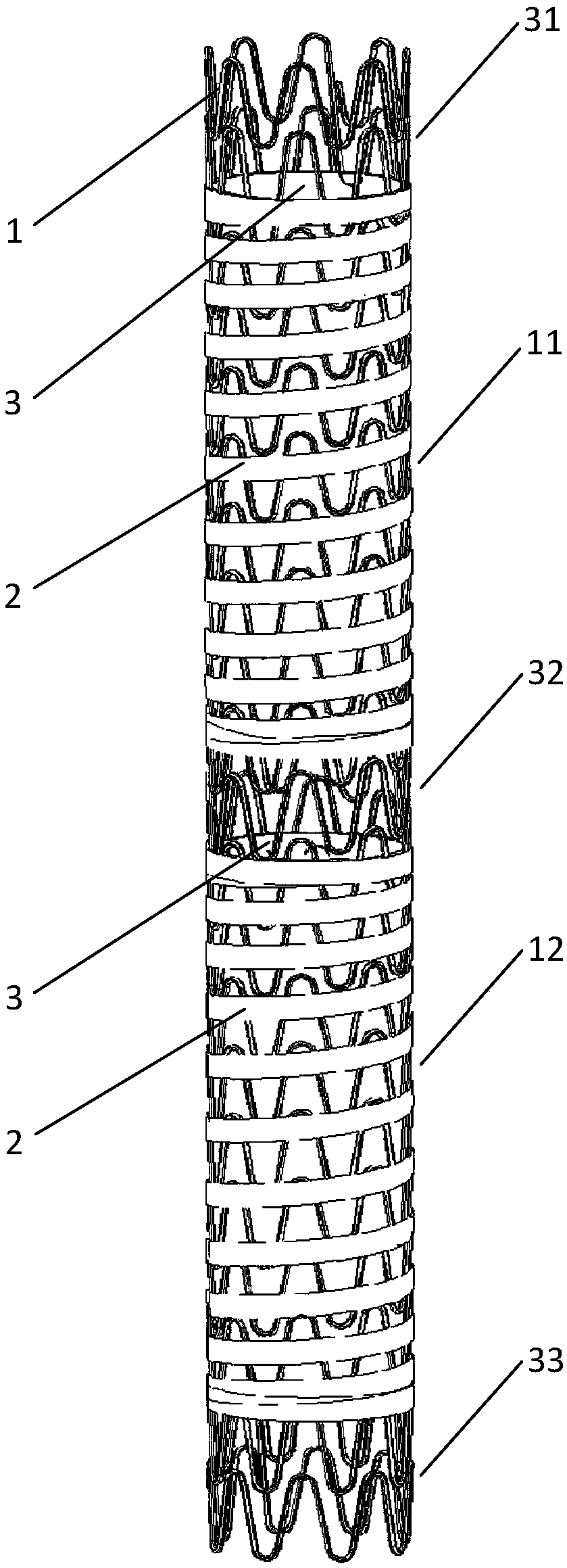
[0044] A drug-coated stent, comprising a stent body 1, an inner covering membrane 2, and an outer membrane 3. The inner covering membrane 2 is closely attached to the inner cavity of the stent body 1, and the inner covering membrane 2 and the stent body 1 fixed connection; the outer membrane 3 is combined with the inner covering membrane 2 by heat sealing, microsurgical suture, etc.; the stent body 1 is provided with an inner covering membrane 2 to form a membrane covering area, and the outer membrane 3 is wound around the The stent body 1 is opposite to the outer surface of the membrane covered area, and the outer membrane 3 firmly wraps the stent body 1 between the inner and outer membranes. On the stent body 1, a bare stent region is formed where the inner membrane 2 is not provided, and the bare stent region and the membrane region are alternately arranged in sequence.

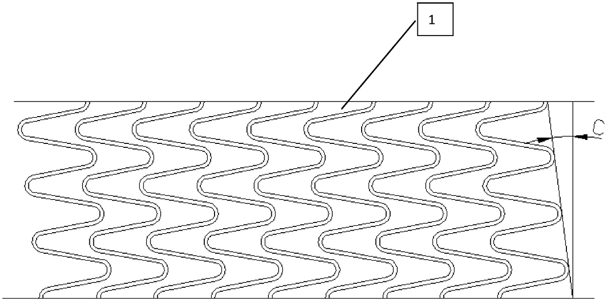
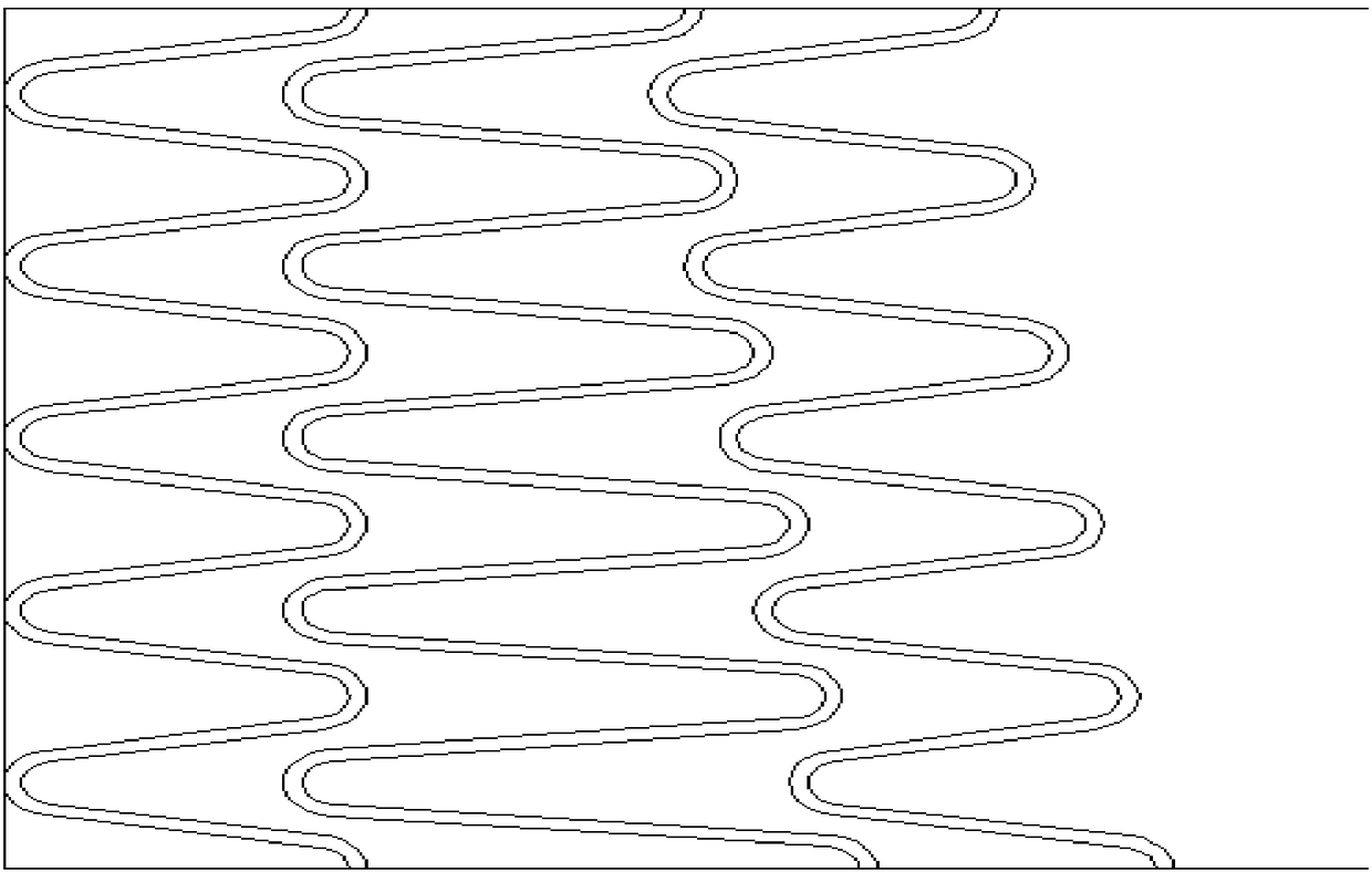
[0045] The stent body 1 is woven from a single wire, the selected single wire has a diameter of 0.1-0.5 mm...

Embodiment 2

[0051] On the basis of Embodiment 1, the metal bracket body is manufactured by laser cutting. Through laser engraving of the pipe, the remaining material and residue are removed, and then through the processes of pickling, electrochemical polishing, cleaning, and drying, a metal bracket body that meets the requirements is formed. Preferably, the tube is a nickel-titanium alloy tube that meets the requirements of the medical implant standard (ASTM F2063), and preferably, the tube has a diameter of 1.65-5.0 mm. In this example, the structure and process of the covered stent covered by the medicine are the same as those in example 1. After testing, the radial support force of the laser-cut stent body before covering is 0.62-1.86N / mm, and the overall radial support force of the coated stent graft is 0.71-1.95N / mm after covering, which improves the stent Support strength to the vascular cavity.

Embodiment 3

[0053] Due to the lumen isolation principle of the coated stents, the endothelialization process of the stent is largely restricted, leading to a high risk of late restenosis. In view of the above shortcomings, under the premise of not affecting the lumen isolation principle of the coated stent graft, a through hole 5 with a certain diameter is added to the inner membrane 2 such as Image 6 As shown, the entrance for the growth of endothelial cells is fully provided, and the process of luminal endothelialization is further accelerated. The method of adding the through hole 5 may be laser drilling. Another way is to retain the bare stent area at both ends, cancel the bare stent area in the middle section of the coated stent graft, and add a large number of through holes with a certain diameter, such as Figure 7 As shown, good results can also be achieved. The above method of adding the through hole 5 may be laser drilling.


PUM


Abstract

The invention discloses a medicine-applying filming support. The medicine-applying filming support comprises a supporting body, an inner film and an outer film. The inner film is closely attached to an inner cavity of the support body and fixedly connected with the support body; filming areas are formed in the portions, provided with the inner film, of the support body, the outer film is wound around the support body relative to the outer surface of the filming areas, and the support body is firmly wrapped by the outer film in the middle of the inner film and the outer film; bare support areas are formed in the portions, not provided with the inner film, of the support body, and the bare support areas and the filming areas are sequentially and alternately arranged. According to the medicine-applying filming support, the bending performance of the filming support is improved through the filming mode; meanwhile, the wall attaching performance and the stability of the filming support are improved, and the problem that the filming support is prone to shifting is effectively solved. The inner cavity of the film is coated with anticoagulation medicine, and thrombosis is reduced; the whole outer wall of the filming support is loaded with medicine for inhibiting smooth-muscle-cell proliferation and migration, and restenosis of the inside and the two ends of the postoperation support cavity is effectively prevented.

Description

Technical field [0001] The invention relates to the field of medical devices, in particular to a drug-coated membrane stent. Background technique [0002] Since parieli took the lead in successfully treating abdominal aortic aneurysms with a drug-applied stent graft in 1991, the drug-applied stent graft has been widely used in aortic diseases. With the development of peripheral vascular intervention techniques, small-caliber peripheral application There are also more clinical reports about the use of stent grafts in clinical practice. [0003] The drug-coated stent graft is composed of an ordinary bare metal stent covered with a special polymer membrane material, which is an effective combination of the supporting physical and chemical properties of the bare metal stent and the unique properties of the membrane material. In the treatment of peripheral arterial occlusive or stenotic diseases, the physical barrier function of the covering material is used to limit the proliferation ...

Claims


Application Information

Patent Timeline
Patent Type & Authority Patents(China)
IPC IPC(8): A61F2/07A61M31/00
CPCA61F2/07A61M31/00A61M2210/12
Inventor 赵中覃元平李峥高飞李秋华
Owner ZHEJIANG BELONGS TO A MEDICAL INSTR
Why Eureka
Try Eureka