Canine mesenchymal stem cell bank and construction method thereof

A kind of mesenchymal stem cell and construction method technology, applied in the field of umbilical cord mesenchymal stem cell bank and its construction, canine placenta, can solve the time-consuming and laborious technical requirements and other problems, achieve the effect of saving operation process time and full of application prospects

Inactive Publication Date: 2017-05-24
天津百恩生物科技有限公司
View PDF5 Cites 14 Cited by
  • Summary
  • Abstract
  • Description
  • Claims
  • Application Information

AI Technical Summary

Problems solved by technology

[0006] Isolation of mesenchymal stem cells by enzymatic digestion and tissue attachment method, easy to mix with other adherent cells, such as macrophages and endothelial cells, on...

Method used


Image

  • Canine mesenchymal stem cell bank and construction method thereof
  • Canine mesenchymal stem cell bank and construction method thereof
  • Canine mesenchymal stem cell bank and construction method thereof

Examples


Embodiment 1

[0037] Embodiment 1: (acquisition of placenta and umbilical cord tissue)

[0038] Screen the donor dogs (ancient shepherd dogs) and select those who pass the physical examination;

[0039] The placenta and umbilical cord of canine animals were taken by cesarean section, and the placental blood was extracted, and the virus detection kit (Shanghai Kuailing Biological Co., Ltd.) was used to infect canine coronavirus, canine distemper virus, rabies virus, canine parvovirus, canine brucellosis, etc. disease detection;

[0040] After passing the test, rinse with phosphate buffered saline (pH 7.3) containing gentamicin and ciprofloxacin to remove residual blood and soak the placental umbilical cord tissue for 15 minutes;

[0041] Cut the placental umbilical cord tissue to about 1-2cm with a sterile knife 3 The small pieces were washed again with phosphate buffer to remove residual blood;

Embodiment 2

[0042] Embodiment 2: (concentration and proportioning screening of mixed digestive juice)

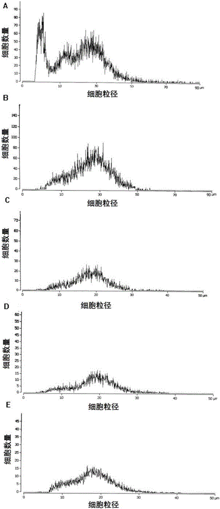
[0043] Divide the placental umbilical cord tissue into 8 parts, add collagenase type I and trypsin (as shown in Table 1) in different proportions and different concentrations for digestion, and dilute and mix the digestive juice with phosphate buffer according to the following proportions, and the volume of the digestive juice is tissue 1 / 2 of the volume, put the container on a constant temperature magnetic stirrer to digest for 30 minutes, the speed is 90-120 rpm, and the temperature is 37 degrees;

[0044] Table 1: Proportion of mixed digestive juice

[0045] group Type I collagenase concentration trypsin concentration volume ratio 1 0.2% 0.5% 1:1 2 0.2% 0.5% 2:1 3 0.2% 0.2% 1:1 4 0.2% 0.2% 2:1 5 0.5% 0.2% 1:1 6 0.5% 0.2% 2:1 7 0.5% 0.5% 1:1 8 0.5% 0.5% 2:1

[0046] After the resulting digestion mixture was p...

Embodiment 3

[0050] Embodiment 3: the comparison of digestion method of the present invention and traditional enzyme digestion method

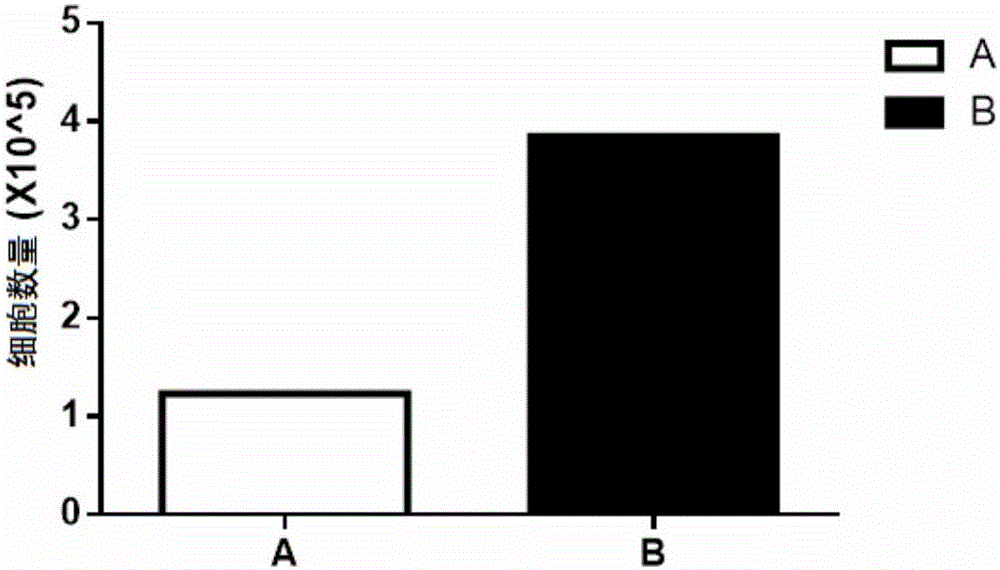
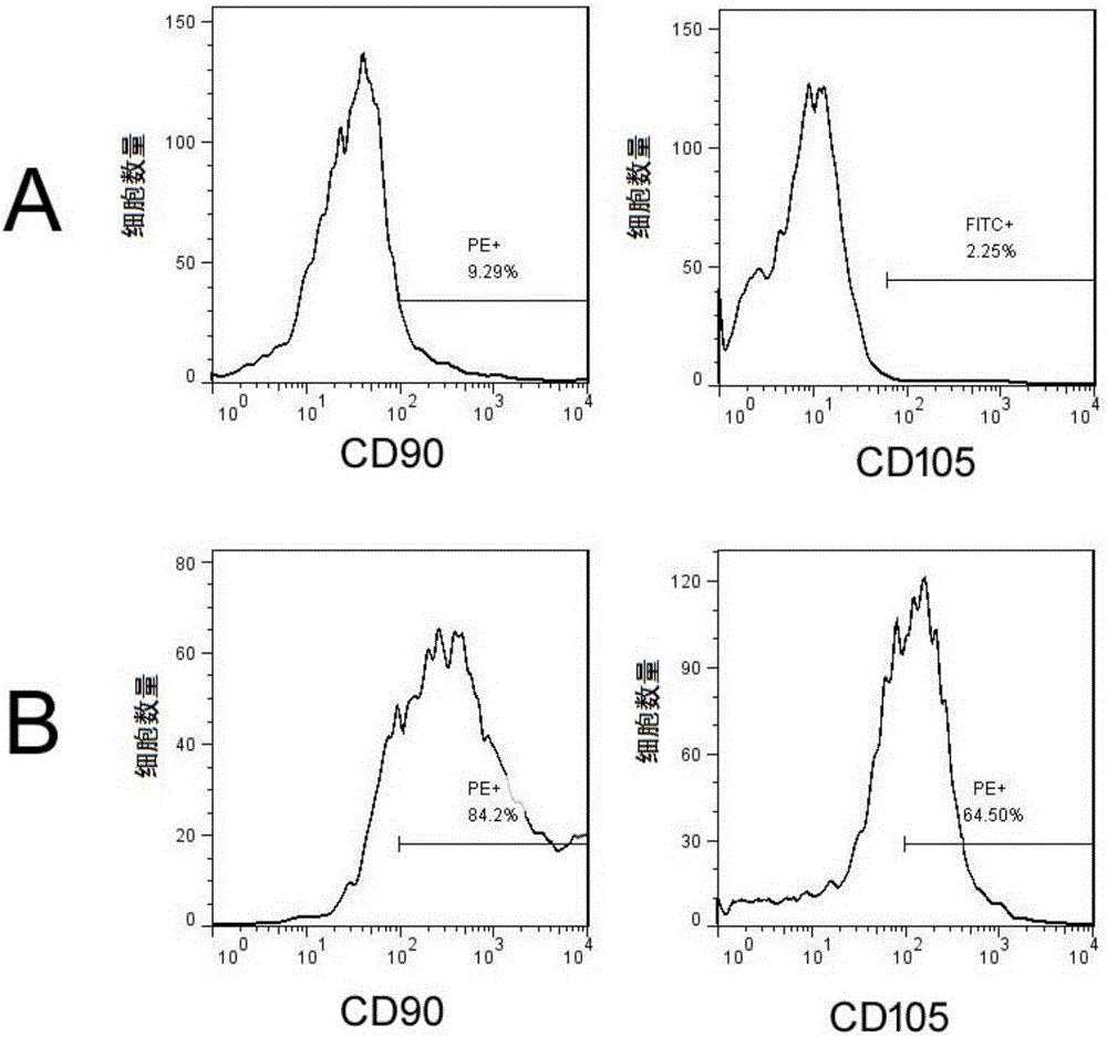
[0051]The digestion method of the present invention: the tissue is put into the container of the digestive juice of type I collagenase (0.5%) and trypsin (0.5%) with the ratio of 1:1, and the container is placed on a constant temperature magnetic stirrer to digest for 30-40 Minutes, temperature 37 degrees; pass the digestion mixture through a cell strainer, add the filtrate to the container for re-digestion, wash the cell strainer with D / F12 medium containing 10-15% dog serum to obtain a suspension of mesenchymal stem cells ; Repeat the digestion 3-5 times, collect the mesenchymal stem cell suspension, centrifuge at 2000 rpm for 10 min, discard the supernatant, and resuspend in D / F-12 medium containing 10-15% dog serum.

[0052] Traditional enzymatic digestion method: put the tissue into the type I collagenase solution with a concentration of 0.6% (w / v), p...


PUM


Abstract

The invention discloses a canine mesenchymal stem cell bank and a construction method thereof. The construction method comprises the following steps: 1, collecting and detecting placentas and umbilical cords; 2, carrying out enzyme digestion on placenta and umbilical cord tissues; 3, collecting cells by using a specially-designed cell filter with multi-mesh sieves; 4, digesting and filtering the cells multiple times, and collecting cells with the particle size of about 10-25 [mu]m; 5, culturing the cells, and identifying the cells; and 6, carrying out liquid nitrogen refrigeration detection, preserving qualified mesenchymal stem cells, and establishing cell information archive to construct the canine placenta and umbilical cord mesenchymal stem cell bank. The mesenchymal stem cells obtained through the method have high activity, can be preserved for a long term, and are simple to operate. The mesenchymal stem cell bank is applied to the treatment of relevant diseases of species relationship similar canoidea.

Description

technical field [0001] The invention relates to a stem cell bank and a construction method thereof, in particular to a canine placenta and umbilical cord mesenchymal stem cell bank and a construction method thereof. [0002] technical background: [0003] Mesenchymal stem cells (MSCs) are a type of stem cells with two important characteristics of stem cells: strong self-proliferation ability and multi-differentiation potential. It can also be obtained from adipose tissue, fetal liver, umbilical cord blood and mobilized peripheral blood, fetal lung, placenta, umbilical cord, dental pulp, synovium, periodontal ligament, endometrium, trabecular bone and compact bone, etc. Isolation and culture of MSCs. [0004] A large number of studies have shown that under appropriate culture conditions, MSCs can differentiate into mesoderm, endoderm and even ectoderm cells, and MSCs can secrete a variety of growth factors and immune protection factors, some of which are important in cell and...

Claims


Application Information

Patent Timeline
IPC IPC(8): C40B50/06C40B40/02C12N5/0775C12N5/073A01N1/02
CPCC40B50/06A01N1/0221C12N5/0605C12N5/0665C12N2509/00C40B40/02
Inventor 梁璐王斌赵宝娜
Owner 天津百恩生物科技有限公司
Why Eureka
Try Eureka