Method for inducing embryonic stem cell to be differentiated into corneal endothelial cells and inducing culture medium

A technology for inducing culture medium and embryonic stem cells, applied in the fields of tissue engineering and ophthalmology, it can solve the problems of cumbersome and long feeder layer co-cultivation method, and achieve the effects of regular morphology, high biological safety and obvious characteristics.

Inactive Publication Date: 2017-05-31
SHANDONG EYE INST
View PDF4 Cites 6 Cited by
  • Summary
  • Abstract
  • Description
  • Claims
  • Application Information

AI Technical Summary

Problems solved by technology

However, the feeder layer co-culture method is cumbersome and takes a long time, and the in vitro process needs to be as simple and fast as possible to better meet future clinical applications.

Method used


Image

  • Method for inducing embryonic stem cell to be differentiated into corneal endothelial cells and inducing culture medium
  • Method for inducing embryonic stem cell to be differentiated into corneal endothelial cells and inducing culture medium
  • Method for inducing embryonic stem cell to be differentiated into corneal endothelial cells and inducing culture medium

Examples


preparation example Construction

[0058] In the present invention, the preparation method of the pre-laid Matrigel preferably comprises the following steps:

[0059] Matrigel was diluted with DMEM / F12 medium and plated;

[0060] Place the plated Petri dish in a sterile environment with a temperature of 37°C and a CO2 volume concentration of 5% for 1-2 hours to gel;

[0061] The gelled DMEM / F12 medium was replaced with mTeSR1 medium to obtain a medium pre-coated with Matrigel. The source of the mTeSR1 medium is preferably purchased from Stem Cell Company.

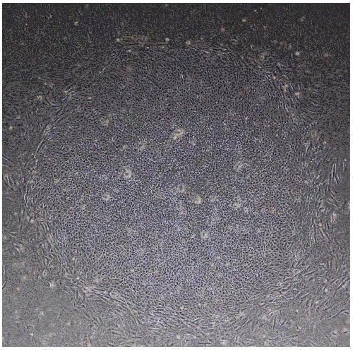
[0062] In the present invention, the time for the basal culture is 3-4 days. After basal culture, human embryonic stem cells reached about 20-100 cells / clones under an inverted phase-contrast microscope, and the clones were uniform in size. The purpose of the basal culture is to grow the stem cell clones to a size that meets the induction conditions.

[0063] To obtain the human embryonic stem cells of the basal culture, the present invention inoculates ...

Embodiment 1

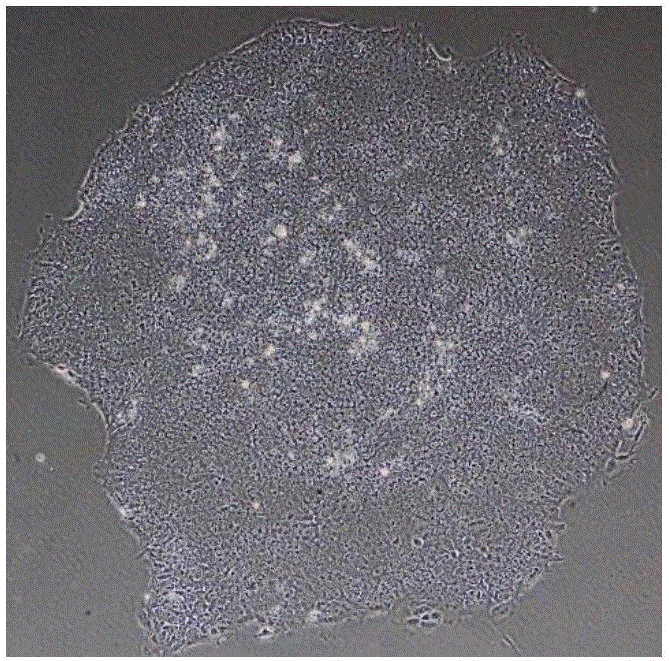
[0078] Culture of human embryonic stem cells: take an aliquoted Matrigel matrigel (purchased from BD Company, product number 354277), and melt on ice at 4°C. Pre-cool pipette tips, Petri dishes, and centrifuge tubes. Use 25ml of cold DMEM / F12 medium (purchased from Invitrogen, product number 11330) to dilute Matrigel to a concentration of 1-2%, and then add 1-2ml to each 60mm diameter petri dish for coating, until the Matrigel is completely After the culture surface was covered, the culture dish was moved into a 37°C, 5% CO2 incubator, and replaced with fresh human embryonic stem cell medium mTeSR1 (purchased from Stem Cell Company, Cat. No. 05850) after 1 hour for future use. When human embryonic stem cells are cultured to a good morphological state (such as figure 1 Shown) Use 3ml of warm DMEM / F12 medium to wash twice, add about 1-1.5ml of the digestive solution Accutase (purchased from Sigma, Cat. 2 Digest in the incubator for 4-7 minutes, observe the edge of the clone un...

Embodiment 4

[0084] Immunofluorescence detection:

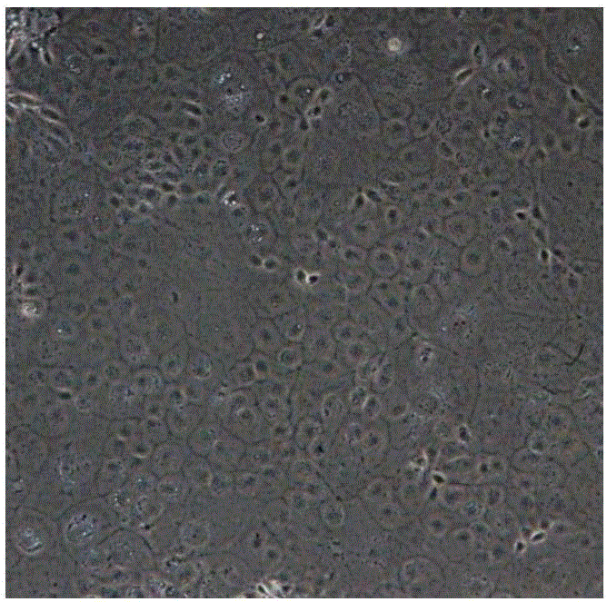
[0085] 1. 4% paraformaldehyde to fix the cells for 15 minutes; 2. Wash 3 times with PBS; 3. 0.1% TritonX-100 permeabilization for 15 minutes (this step can be omitted for membrane antigen); 4. Add 5% BSA to block at room temperature for 1 hour; 5. Add the corresponding primary antibody and incubate at room temperature for 2 hours or overnight at 4°C; 6. Add fluorescent secondary antibodies corresponding to the source of the primary antibody, and incubate at room temperature for 1 hour; 7. DAPI staining and take pictures under a fluorescent microscope. Fluorescent immunographs of cell markers such as Figure 5 and Figure 6 shown. Figure 5 Expression of corneal endothelial fluid pump function marker Na for differentiated cells + -K + - Immunofluorescence pictures of ATPase markers; Figure 6 It is a positive control picture, which is normal human corneal endothelial cells expressing the corneal endothelial cell fluid pump function ma...


PUM


Abstract

The invention provides an inducing culture medium for inducing to produce corneal endothelial cells. The inducing culture medium is characterized by taking a DMEM (Dulbecco Modified Eagle Medium)/F12 culture medium as a basic culture medium, and is prepared from the following components: beta-mercaptoethanol of which the molar concentration is 0.05 to 0.15 mmol/L, glutamine of which the molar concentration is 0.05 to 0.15 mmol/L, bFGF (Basic Fibroblast Growth Factors) of which the mass concentration is 4 to 8 ng/ml, NEAA (Non Essential Amino Acid) of which the molar concentration is 0.05 to 0.15 mmol/L, KSR (Knockout Serum Replacement) of which the volume concentration is 18 to 25 percent and RA of which the molar concentration is 0.5 to 2 mu mol/L. The invention also provides a method for inducing an embryonic stem cell to be differentiated into the corneal endothelial cells, wherein the corneal endothelial cells which are obtained through inducing are obvious in morphology rule feathers. The method does not involve a feeder layer, so that other animal origin pollution is avoided, and the biological safety is high; the corneal endothelial cells are used as corneal endothelium seed cells in tissue engineering, so that a qualified material source is provided for clinical corneal endothelium transplantation.

Description

technical field [0001] The invention belongs to the fields of tissue engineering and ophthalmology, and specifically relates to a method for inducing embryonic stem cells to differentiate into corneal endothelial cells and an induction medium. Background technique [0002] The corneal endothelium is the innermost single layer of cells in the cornea that maintains the transparency of the cornea through its active fluid pump function. The proliferation ability of human corneal endothelial cells is extremely limited, and the damage is mainly repaired by the expansion and migration of peripheral cells in the damaged area. Various reasons such as Fuch corneal endothelial dystrophy, trauma and surgery can cause damage to corneal endothelial cells. When the cell density is lower than 500-1500 / mm 2 Corneal endothelial function decompensation occurs from time to time, resulting in corneal edema and turbidity, and bullous keratopathy. Cornea and its component transplantation is a bri...

Claims


Application Information

Patent Timeline
Patent Type & Authority Applications(China)
IPC IPC(8): C12N5/079
CPCC12N5/0621C12N2500/32C12N2500/33C12N2500/38C12N2500/44C12N2501/115C12N2506/02
Inventor 周庆军段豪云史伟云王瑶
Owner SHANDONG EYE INST
Why Eureka
Try Eureka