A kit for breast cancer detection based on liquid biopsy

A technology of liquid biopsy and reagent kit, which is applied in the determination/inspection of microorganisms, biochemical equipment and methods, etc., which can solve problems such as difficult to distinguish, different amounts of antigens, weak fluorescence intensity, etc., and achieve good observation and high enrichment efficiency Effect

Active Publication Date: 2020-03-31
上海美吉医学检验有限公司
View PDF6 Cites 0 Cited by
  • Summary
  • Abstract
  • Description
  • Claims
  • Application Information

AI Technical Summary

Problems solved by technology

However, this method can only specifically identify the rare cells through a single monoclonal antibody, which is likely to cause missed detection
At the same time, due to the heterogeneity of cells, the amount of antigen expressed by each cell is different, resulting in sometimes very weak fluorescence intensity, which is difficult to distinguish under a fluorescence microscope

Method used


Image

  • A kit for breast cancer detection based on liquid biopsy
  • A kit for breast cancer detection based on liquid biopsy
  • A kit for breast cancer detection based on liquid biopsy

Examples


Embodiment 1

[0057] Example 1 enrichment of target cells in peripheral blood

[0058] (1) Centrifuge peripheral blood to remove plasma protein: 8.5mL peripheral blood was centrifuged at 800g in a horizontal centrifuge for 7min at room temperature, and the supernatant was discarded.

[0059] (2) Add 5-6 mL of PBS buffer solution and 3 mL of lymphocyte separation solution to the plasma in step (1), centrifuge at 450 g in a centrifuge for 7 minutes at room temperature. After centrifugation, it is divided into three layers. The red bottom layer is the erythrocyte layer, the slightly white middle layer is mainly white blood cells and CTC, etc., and the yellow upper layer is plasma. Absorb all the liquid above the erythrocyte layer and remove the bottom erythrocyte layer.

[0060] (3) Add 200 mL of immunomagnetic beads with CD45 antibody coupled to the surface dropwise in step (2), incubate on a horizontal shaker to obtain a suspension, set the rotation speed of the horizontal shaker at 120-150 ...

Embodiment 2

[0062] Example 2 Fluorescent staining of enriched target cells

[0063] (1) Enhanced staining pretreatment: Add 2 μL of staining enhancement solution to about 50 μL of enriched target cells, and let stand at room temperature for 10 min. The staining enhancing solution is a PBS buffer solution of SDS or Triton X-100, and the SDS concentration is 0.1 mg / mL.

[0064] (2) Cell surface staining: After diluting CD45-Alexa 5941 μL with 199 μL of PBS buffer, it was added to the cell suspension after the pretreatment in step (1), and then incubated in the dark for 20 minutes. After incubation, add PBS buffer to wash the cell liquid, centrifuge at 950g for 4min, and remove the supernatant to 100μL.

[0065] (3) Cell fixation: transfer and smear the cells in step (2) onto a glass slide, then add the fixative paraformaldehyde, fix for 10 min, and wash the slide twice with PBS, 5 min each time.

[0066] (4) Cell membrane rupture: After the cells were fixed, 200 μL of cell membrane ruptur...

Embodiment 3

[0070] Fluorescence staining detection of embodiment 3 cell lines

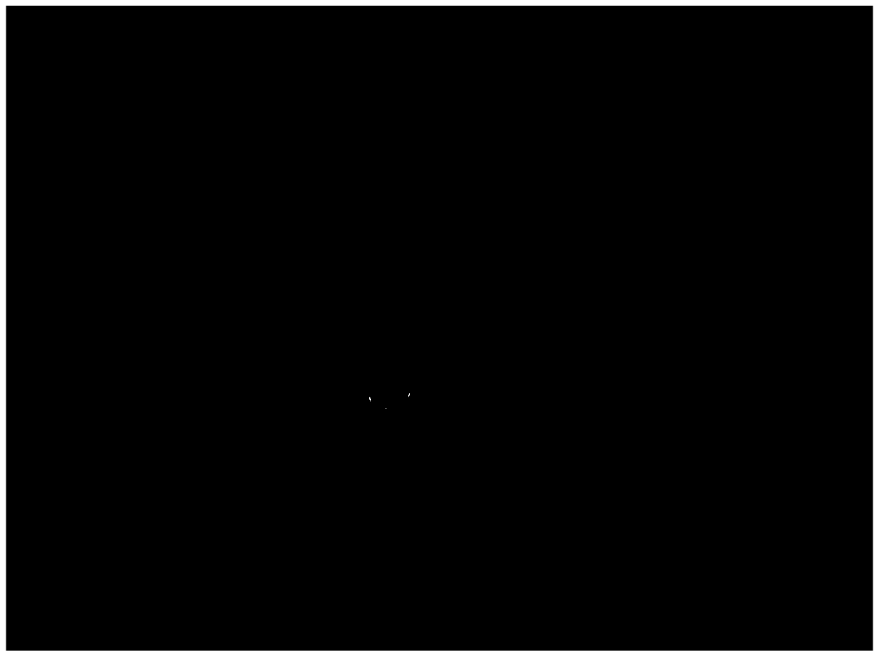
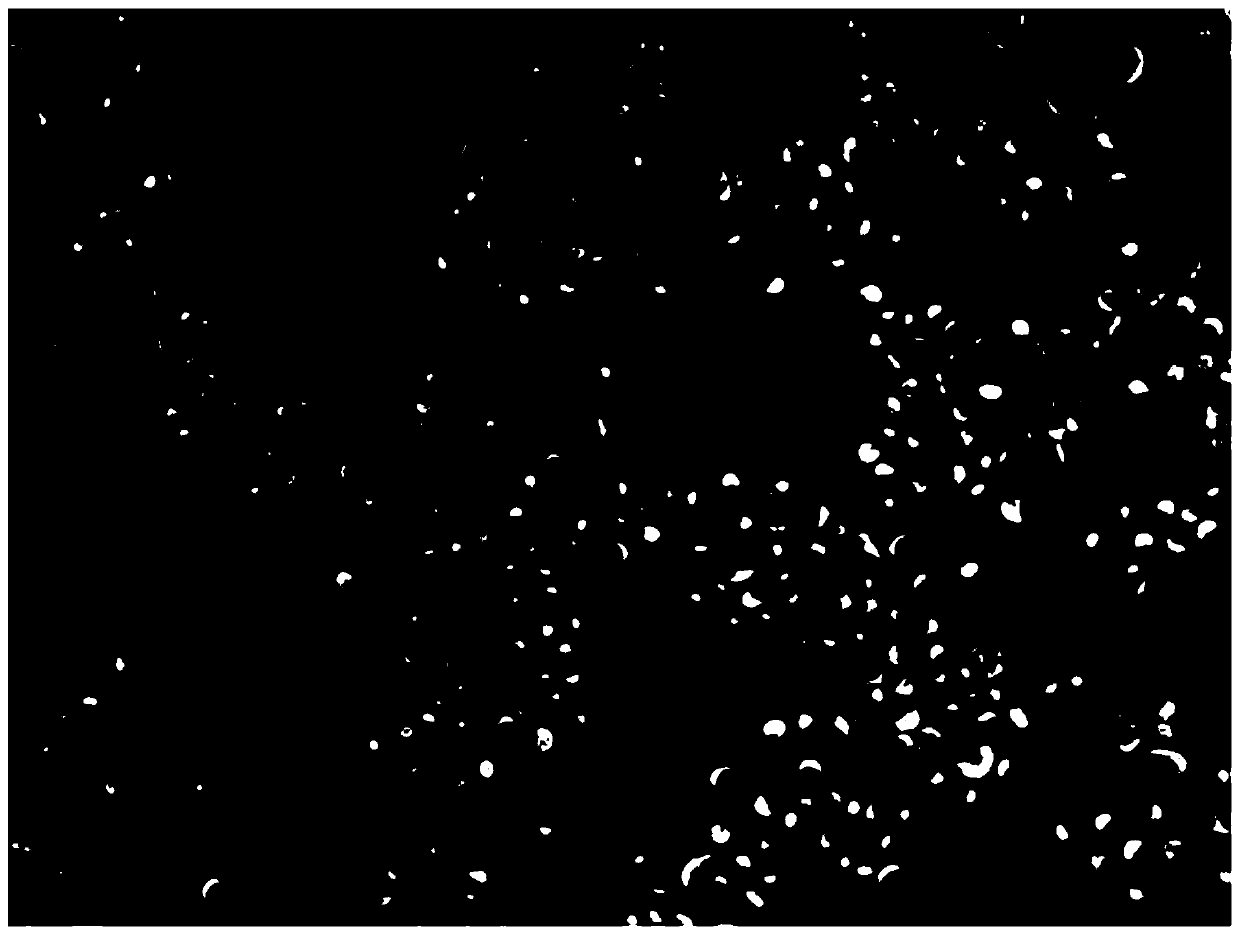
[0071] MCF7 (ERα+ / HER2-) or SKBr3 (ERα- / HER2+) or MDA-MB-231 (ERα- / HER2- / VIM+) cell lines (purchased from the Cell Bank of the Chinese Academy of Sciences) were digested by enzymatic digestion and 10 5 Cells, approximately 50 μL, were subjected to cell fluorescence staining and fluorescence microscope examination according to the steps in Example 2. Microscopic examination conditions are as follows: when the excitation light wavelength is 591nm, Alexa594 emits 618nm red light, and the exposure time is 100ms; when the excitation light wavelength is 650nm, CY5 emits 670nm far-red light (invisible to the naked eye, and the microscope scanning is assigned purple). The time is 100ms; when the excitation light wavelength is 499nm, Alexa488 emits 519nm green light, and the exposure time is 100ms; when the excitation light wavelength is 345nm, DAPI emits 455nm blue light, and the exposure time is 10-20ms, the results ...


PUM


Abstract

The invention provides a reagent kit for detecting breast cancer on the basis of liquid biopsy. The reagent kit comprises staining enhancement solution for enhancing staining effects and specific antibodies with fluorescence staining markers. The specific antibodies include ER alpha antibodies, VIM antibodies, CD45 antibodies and HER2 antibodies; the staining enhancement solution comprises surfactants with the concentration of 0.001-1 mg / mL. The reagent kit has the advantages that target cells can be effectively enriched, whether the target cells come from early-stage patients who suffer from the breast cancer or not can be confirmed, and tumor can be classified; the tumor detection sensitivity can be improved by means of three-tumor-marker detection, and the detection accuracy further can be guaranteed by means of CEP8 detection; the staining effects can be enhanced by the staining enhancement solution, accordingly, the diversified antibodies with the fluorescence staining markers can be combined with the target cells, the target cells can be stained, the good staining effects can be realized, fluorescence is intensive, and boundaries are clear.

Description

technical field [0001] The invention relates to the field of liquid biopsy, in particular to a kit for detecting breast cancer based on liquid biopsy. Background technique [0002] Breast cancer is a disease that seriously threatens women's health. Its incidence rate accounts for 23% of all tumors, ranking first among women, and its mortality rate ranks second among all cancers. According to statistics, in my country, 30-40 breast cancer patients are diagnosed for every 100,000 population every year, and the average age of breast cancer diagnosis is 45-55 years old, which is younger than Western women. Therefore, it is particularly important for the early screening of breast cancer and the detection during the development of the disease. [0003] At present, for the early screening of breast cancer, the method of detecting circulating tumor cells (CTC) based on liquid biopsy has many advantages that traditional detection methods do not have, including non-invasive, fast, an...

Claims


Application Information

Patent Timeline
Patent Type & Authority Patents(China)
IPC IPC(8): C12Q1/6841
CPCC12Q1/6841C12Q2543/10C12Q2563/107
Inventor 陈昌岳段彪张培培张祥林蔡红东李静冯丽娜
Owner 上海美吉医学检验有限公司
Why Eureka
Try Eureka