Adjustable luciferase segmentation fusion protein and preparing method and application thereof

A luciferase and fusion protein technology, applied in the field of molecular biology, can solve the problems of poor real-time performance and differences of dual fluorescence complementary technology, which can only be detected successfully in a few minutes or even a few hours

Active Publication Date: 2017-07-04
SHENZHEN INST OF ADVANCED TECH CHINESE ACAD OF SCI
View PDF4 Cites 12 Cited by
  • Summary
  • Abstract
  • Description
  • Claims
  • Application Information

AI Technical Summary

Problems solved by technology

However, the real-time performance of the double fluorescent complementation technology (BiFC) is poor. It requires that the two fragments can be complementary and reintegrated on the cut surface to form a complete fluorescent protein to restore the activity of the fluorescent protein. Fluorescent co

Method used


Image

  • Adjustable luciferase segmentation fusion protein and preparing method and application thereof
  • Adjustable luciferase segmentation fusion protein and preparing method and application thereof
  • Adjustable luciferase segmentation fusion protein and preparing method and application thereof

Examples


Embodiment 1

[0047] Example 1 Preparation of Gausssia luciferase fusion fragment

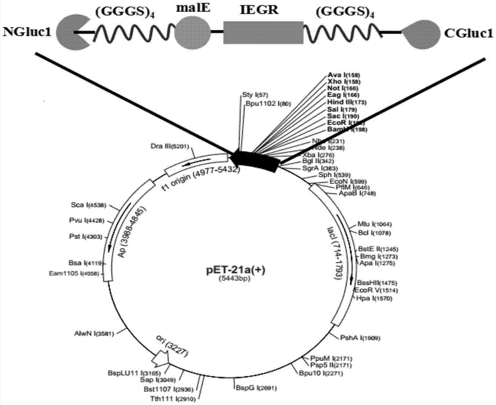
[0048] (1) Select the expression vector of the luciferase fusion gene as pET21a, and then select a suitable insertion site for luciferase, and finally choose to insert an exogenous fragment between Arg93 and Cys94 to form the N fragment NGluc1 (1~ 93aa) and C fragment CGluc1 (94~185aa);

[0049] (2) Using the recombinant vector pET28a-Gluc1 as a template, when designing and amplifying the NGluc1 fragment, introduce BamH Ⅰ and EcoR Ⅰ restriction sites at the 5' end of the primer, and when designing and amplifying the CGluc1 fragment, introduce HindⅢ at the 5' end of the primer and Not I restriction sites, and are designed to introduce flexible peptide chains (GGGGS) 4 To reduce the steric hindrance caused by specific enzyme cleavage;

[0050] The designed primer sequences are as follows:

[0051] Gluc1 upstream primer (BamH Ⅰ):

[0052] 5'-CGCGGATCCATGAAGCCCACCGAGAACAACGAAG-3';

[0053] NGluc1 downstream...

Embodiment 2

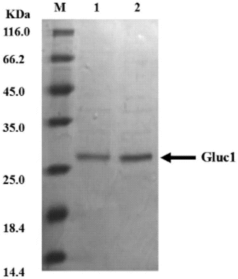
[0069] Embodiment 2 Expression and purification of luciferase fusion body and Gaussia luciferase

[0070] (1) Strain activation: inoculate frozen glycerol bacteria pET21a-NGluc1-malE-CGluc1 and pET28a-Gluc1 into LB liquid medium (containing 100 μg / mL ampicillin) at 37°C and 225 rpm to activate overnight strains;

[0071] (2) Strain fermentation: Inoculate the activated bacterial liquid into fresh LB liquid medium (containing 100 μg / mL ampicillin) at a ratio of 1:100 and cultivate to OD 600 When it reaches 0.4-0.6, it is the logarithmic phase of the growth of the strain;

[0072] (3) Induced expression: add inducer isopropyl-β-D-1-thiogalactoside IPTG (final concentration is 1mM) to induce high-speed expression of luciferase fusion at 28°C, centrifuge at 11000rpm at 4°C Collect the thalli within 10min, discard the supernatant LB medium, add a certain amount of binding buffer (20mM Na 3 PO 4 , 0.5M NaCl, 40mM imidazole, pH 7.4) suspended bacteria;

[0073] (4) Ultrasonic dis...

Embodiment 3

[0075] Example 3 Construction of the dynamic interaction model of luciferase fusion protein

[0076] (1) Realize the ON mode of protein dynamic interaction

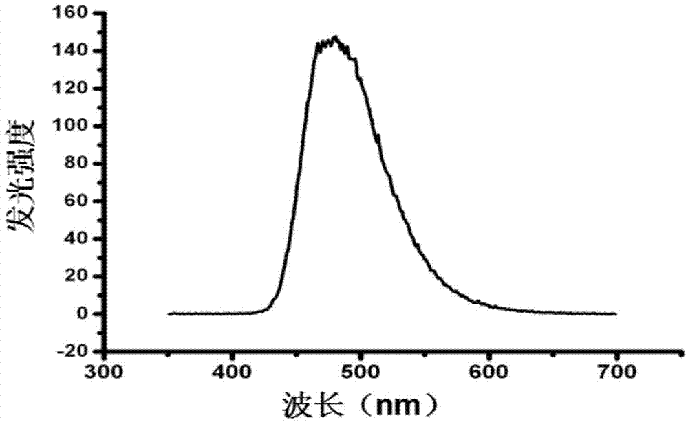
[0077] Add 1×PBS (pH7.4) buffer solution to a 96-well white microtiter plate, add luciferase fusion protein diluted to a certain concentration with buffer solution, and quickly add intestinal Coelenterazine (Coelenterazine) substrate, to ensure that the total volume of the system is 200 μL, after thorough mixing, immediately use a microplate reader to detect the bioluminescent intensity in the system (the emission spectrum of the fluorescein surface is shown in image 3 As shown, the detection wavelength is 480-500nm), and the ON mode of the protein dynamic interaction model is realized. The model was evaluated with Gaussia luciferase as a control group;

[0078] (2) Realize the OFF mode of protein dynamic interaction

[0079] Add a certain concentration of cutting enzyme Factor Xa Protease to the reaction system, mix ...


PUM


Abstract

The invention relates to the technical field of molecular biology, in particular to adjustable luciferase segmentation fusion protein and a preparing method and application thereof. The fusion protein comprises luciferase N-terminal protein, a flexible peptide fragment 1, MBP protein, an active valve, a flexible peptide fragment 2 and luciferase C-terminal protein, wherein luciferase N-terminal protein and luciferase C-terminal protein form luciferase together, and the active valve is a fragment of 4-8 hydrophobic short-peptide amino acids. By means of the activity adjustment mode that a short-peptide chain amino acid sequence is used as the active valve and specific cleavage enzyme is used as a valve key, a novel fluorescence complementation protein switching mode is realized, and the novel fluorescence complementation protein switching mode can be used for evaluating interaction of other protein dynamics such as combination of a ligand and a substrate, interaction capable of generating a new binding site, protein-protein interaction capable of deactivating protein, and protein-protein interaction capable of changing the specificity of a protein active substrate.

Description

technical field [0001] The invention relates to the technical field of molecular biology, in particular to an adjustable luciferase fragment fusion protein, its preparation method and application. Background technique [0002] With the in-depth study of subcellular structure and function, molecular physiology and pathology, and intercellular and intracellular signaling pathways, human beings' understanding of diseases and the essence of life has been traced back to the level of proteins and genes. Many life activities are realized by the combination and dissociation of protein molecules. Various important physiological activities of cells, the response of cells to the external environment and internal environment, etc., are all based on the interaction between proteins. guide the biological activity of cells and form a signal transduction network system. Therefore, an in-depth study of protein interactions is a necessary prerequisite for the recognition and understanding of...

Claims


Application Information

Patent Timeline
IPC IPC(8): C12N9/02C12N15/62C12N15/70C12Q1/66C12Q1/37
CPCC12N9/0069C12Q1/37C12Q1/66C07K2319/50C07K2319/61C07K2319/24
Inventor 金宗文袁静罗擎颖
Owner SHENZHEN INST OF ADVANCED TECH CHINESE ACAD OF SCI
Why Eureka
Try Eureka