Method for quantitatively analyzing nicotine-induced cell DNA damage based on high content technology

A DNA damage and quantitative analysis technology, applied in the analysis of materials, material excitation analysis, material analysis by optical means, etc., can solve the problem of unable to provide intracellular fluorescence focus distribution, damage to cell structure and functional integrity, microscopy technology detection problems such as low throughput, to achieve the effect of simple and fast sample processing, improved detection efficiency and sensitivity, and sensitive and accurate detection results

Inactive Publication Date: 2017-09-12
CHINA NAT TOBACCO QUALITY SUPERVISION & TEST CENT +1
View PDF5 Cites 3 Cited by
  • Summary
  • Abstract
  • Description
  • Claims
  • Application Information

AI Technical Summary

Problems solved by technology

Flow cytometry and Western Blot are cumbersome to operate, and adherent cells need to be enzymatically hydrolyzed into single-cell suspension before detection, which destroys the structural and functional integrity of cells, and the detection throughput is low
ELISA cannot provide the distribution of fluorescent focal points in cells and needs to add other detection proteins to correct the results, while the detection throughput of microscopy technology is low, and the error of human counting is large

Method used


Image

  • Method for quantitatively analyzing nicotine-induced cell DNA damage based on high content technology
  • Method for quantitatively analyzing nicotine-induced cell DNA damage based on high content technology
  • Method for quantitatively analyzing nicotine-induced cell DNA damage based on high content technology

Examples


Embodiment 1

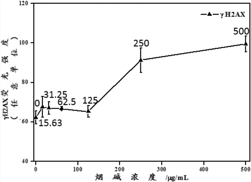
[0064] When the metabolic activation system rat liver S9 was not added to the cell poisoning solution, the γH2AX induced by nicotine after 24h exposure was measured.

[0065] A549 cells in the logarithmic growth phase were collected and planted in 96-well plates at 10,000 cells per well in RPMI-1640 medium containing 10% FBS and 2mmoL / LL-glutamine at 37°C, 5% CO 2 Incubate in the incubator for 24h.

[0066] Aspirate and discard the cell culture fluid after culturing for 24 h, and use cell culture fluid containing 0, 15.63, 31.25, 62.5, 125, 250 and 500 μg / mL nicotine respectively (also containing 10% FBS and 2mmoL / LL-glutamine) RPMI-1640 culture medium) was used as the cell poisoning solution, and the cells were continued to be cultured for 24 h.

[0067] Discard the cell poisoning solution after exposure, add 100 μL PBS to each well and wash twice for at least 5 minutes each time; then add 50 μL 4% paraformaldehyde solution to each well and fix at room temperature for 15 min...

Embodiment 2

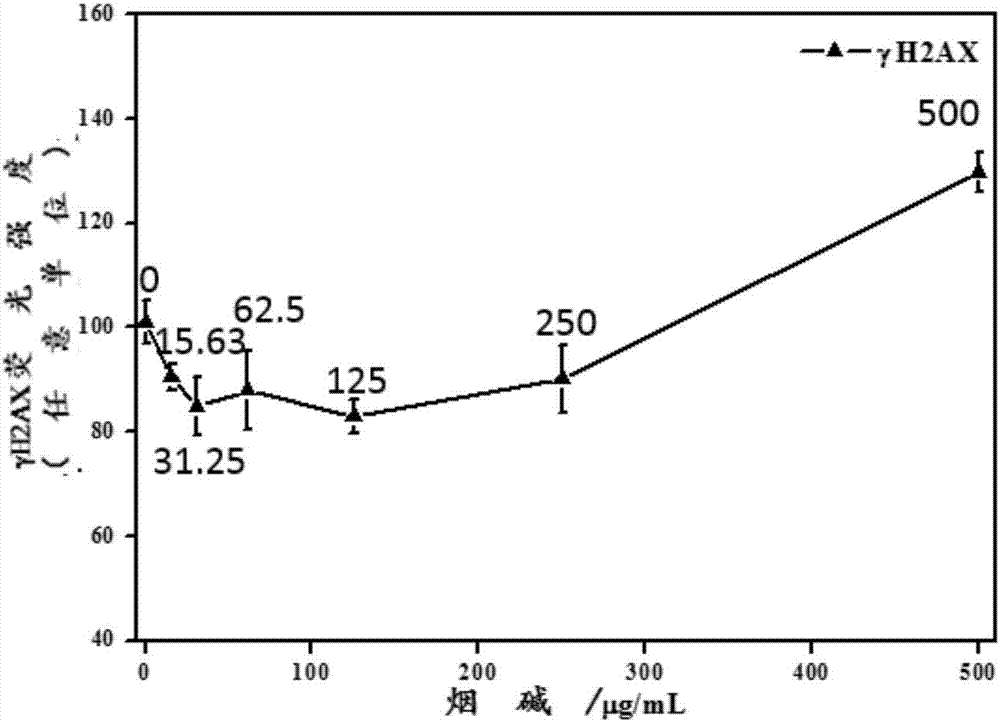
[0072] When the metabolic activation system rat liver S9 was added to the cell poisoning liquid, the γH2AX induced by nicotine after 24h exposure was measured.

[0073] The experimental process was carried out as described in Example 1, the only difference being that, in addition to the cell culture medium and nicotine, the cell poisoning solution was mixed with 10% S9 mixture, so that the cell poisoning solution contained 1% S9 mixture.

[0074] image 3 Shown is the dose-effect relationship curve of γH2AX produced by A549 cells induced by different concentrations of nicotine after 1% S9 was added to the cell poisoning solution 24 hours after exposure. As can be seen from the figure, with the increase of the exposure concentration, the γH2AX produced in the cells first decreased and then increased.

[0075] The addition of S9 to the cell poisoning solution can enhance the metabolic transformation of nicotine in vitro, so the addition of the in vitro metabolic activation syst...

Embodiment 3

[0077] When the metabolic activation system rat liver S9 was not added to the cell poisoning solution, the γH2AX induced by 500 μg / mL nicotine were measured after 1, 2, 4, 8, 12 and 24 hours of exposure, respectively.

[0078] The experimental process was carried out as described in Example 1, with the only difference that the concentration of nicotine in the cell-infected solution was fixed at 500 μg / mL, and the exposure time was 1, 2, 4, 8, 12 and 24 hours, respectively.

[0079] Figure 4Shown is the time-effect relationship curve of γH2AX produced by A549 cells induced by 500 μg / mL nicotine at 1, 2, 4, 8, 12 and 24 hours after exposure. It can be seen from the figure that as the exposure time increases, the γH2AX produced by A549 cells gradually increases, showing a significant time-effect relationship.


PUM


Abstract

The invention discloses a method for quantitatively analyzing nicotine-induced cell DNA damage based on the high content technology. The method includes the steps of firstly, culturing cells; infecting the cells; thirdly, performing immunofluorescent labeling of gamma H2AX; fourthly, performing high-content-technology quantitative analysis. The method has the advantages that a high-content imaging system is used to perform automatic imaging, DNA double-strand break marker gamma H2AX protein generated by nicotine induction is quantitatively analyzed, direct and fast cell detection is achieved, and sample processing convenience is achieved; the high-resolution imaging allows the distribution of the gamma H2AX in each cell nucleus to be able to be observed directly and image data to be convenient to store and reanalyze, the nicotine-induced gamma H2AX in each cell nucleus can be quantitatively analyzed, and the detection result is sensitive and accurate.

Description

technical field [0001] The invention belongs to the technical field of in vitro detection of DNA damage, and more specifically relates to an in vitro quantitative analysis method for cellular DNA damage caused by nicotine. Background technique [0002] Nicotine is an addictive substance in tobacco products and smoke, which can cause cardiovascular disease and reproductive toxicity. Although it is generally believed that nicotine is not a carcinogen, it may be a carcinogenic enhancer. Micronucleated cells in the peripheral blood cells of rats exposed to nicotine significantly increased; in vitro experiments showed that nicotine can cause DNA strand breaks through oxidative stress. Currently, the World Health Organization Framework Convention on Tobacco Control (FCTC) lists nicotine as one of the priority pollutants in smoke control. In addition, nicotine is a precursor of nitrosamines such as N-nitrosonornicotine, which may be converted into carcinogenic nitrosamines in the...

Claims


Application Information

Patent Timeline
Patent Type & Authority Applications(China)
IPC IPC(8): G01N21/64
CPCG01N21/6428
Inventor 胡清源张森侯宏卫陈欢王安刘勇
Owner CHINA NAT TOBACCO QUALITY SUPERVISION & TEST CENT
Why Eureka
Try Eureka