Preparation method of natural soft tissue decellularization matrix
A decellularized matrix and soft tissue technology, applied in the fields of biomedicine, regenerative medicine, and clinical medicine, can solve the problems of matrix composition and three-dimensional structure destruction, time-consuming, serious reagent residues, etc., to weaken the interaction, shorten the time, The effect of improving cell removal efficiency
- Summary
- Abstract
- Description
- Claims
- Application Information
AI Technical Summary
Problems solved by technology
Method used
Image
Examples
Embodiment 1
[0061] Decellularization of natural soft tissues to obtain acellular matrix, taking the submucosa of the pig small intestine as an example, includes the following steps:
[0062] Material: Take the jejunum section of the pig small intestine and clean it with 0.9wt% physiological saline;
[0063] Pretreatment: Carefully peel and remove the serosal layer and base layer in the intestinal wall of the pig small intestine, scrape off the mucosal layer to obtain the submucosa of the pig small intestine, and clean it with 0.9wt% physiological saline;
[0064] Disinfection: soak in a solution containing 0.02wt% peroxyacetic acid and 5% ethanol for 1h, and rinse with 0.9wt% physiological saline;
[0065] Degreasing: Put the sterilized submucosa of the pig small intestine into a degreasing reagent for 4 hours. The degreasing reagent is chloroform. The total volume of the degreasing reagent is 30 times the volume of the submucosa of the pig small intestine;
[0066] Enzyme treatment: Put the defatt...
Embodiment 5
[0081] With reference to the national standard YY / T 0606.25-2014, the undecellularized submucosa of the pig small intestine and the submucosa of the pig small intestine after the decellularization of Examples 2-1 to 4-3 were tested for the amount of residual DNA. The DNA residue in the submucosa of the pig small intestine treated by the decellularization process is 696.53ng / mg. The test results of Examples 2-1 to 4-3 are shown in Table 4. After the above decellularization process, the submucosal matrix of the pig small intestine The amount of residual DNA in the slab is significantly reduced.
[0082] Table 4 DNA residues in the submucosa of pig small intestine with different process parameters
[0083]
Embodiment 6
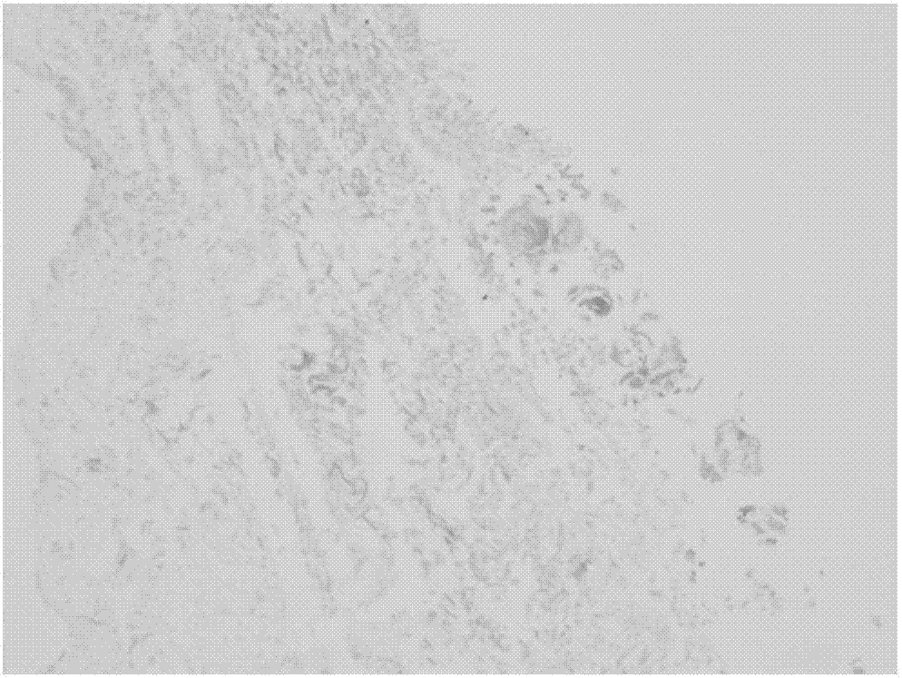
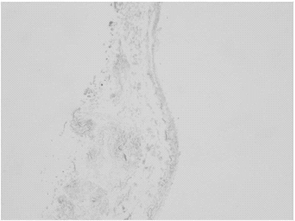
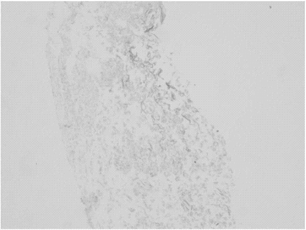
[0085] Perform HE staining on the porcine small intestinal submucosal matrix after decellularization in Example 1 and Examples 4-1, 4-2, and 4-3, such as Figure 1A , Figure 1B , Figure 1C , Figure 1D As shown, the staining results show that there are basically no cells in the submucosa of the pig small intestine treated in Example 1 and Examples 4-1, 4-2, and 4-3, and no significant difference is observed under the microscope. It shows that this process can effectively remove the cells in the submucosal matrix of the pig small intestine and reduce the immunogenicity of the matrix.
PUM
 Abstract
Description
Claims
Application Information
 - R&D
- Intellectual Property
- Life Sciences
- Materials
- Tech Scout
- Unparalleled Data Quality
- Higher Quality Content
- 60% Fewer Hallucinations
Browse by: Latest US Patents, China's latest patents, Technical Efficacy Thesaurus, Application Domain, Technology Topic, Popular Technical Reports.
© 2025 PatSnap. All rights reserved.Legal|Privacy policy|Modern Slavery Act Transparency Statement|Sitemap|About US| Contact US: help@patsnap.com


