PG (pepsinogen) and H.pylori antibody detection method and kit
A technology of pepsinogen and Helicobacter pylori, applied in the field of biomedicine, can solve the problems of inaccurate quantification, high detection sensitivity, and complicated operation process, and achieve rapid and highly sensitive determination, high sensitivity, and strong specificity
- Summary
- Abstract
- Description
- Claims
- Application Information
AI Technical Summary
Problems solved by technology
Method used
Image

Examples
preparation example Construction
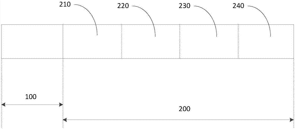
[0044] The general preparation method for detecting pepsinogen Ⅰ, pepsinogen Ⅱ and anti-Helicobacter pylori antibody lateral flow chromatography detection reagent comprises the following steps:
[0045] (1) Preparation of carboxyl-modified fluorescent microsphere-labeled probes
[0046] Mix styrene and methyl methacrylate at a ratio of 1:1, add 1% rare earth complex Eu(TTA)3Phen or 0.5% CdSe / ZnS quantum dots, and ultrasonically mix to obtain liquid a. 0.05% carboxylated polyvinyl alcohol and 0.05% sodium bicarbonate were dissolved in water to obtain liquid b. Add liquid a to liquid b and ultrasonicate for 15 minutes, blow nitrogen for 30 minutes, stir to remove oxygen, and then heat to 80 degrees. Add 0.01-0.1% potassium persulfate and react for 12 hours to obtain polymer fluorescent microspheres, which are filtered, centrifuged and washed with deionized water to obtain purified functionalized fluorescent microspheres.
[0047] After activating the carboxyl groups on the sur...
Embodiment 1
[0060] Example 1 Preparation of Lateral Flow Detection Reagent for Gastric Cancer Screening
[0061] (1) Preparation of fluorescent microsphere-labeled pepsinogen Ⅰ and pepsinogen Ⅱ-labeled antibodies, Helicobacter pylori-labeled antigen and mouse IgG
[0062] Mix styrene and methyl methacrylate at a ratio of 1:1, add 1% rare earth complex Eu(TTA)3Phen or 0.5% CdSe / ZnS quantum dots, and ultrasonically mix to obtain liquid a. 0.05% carboxylated polyvinyl alcohol and 0.05% sodium bicarbonate were dissolved in water to obtain liquid b. Add liquid a to liquid b and ultrasonicate for 15 minutes, blow nitrogen for 30 minutes, stir to remove oxygen, and then heat to 80 degrees. Add 0.01-0.1% potassium persulfate and react for 12 hours to obtain polymer fluorescent microspheres, which are filtered, centrifuged and washed with deionized water to obtain purified functionalized fluorescent microspheres.
[0063] Take 20 mg of the above-mentioned carboxyl-modified fluorescent microspher...
Embodiment 2
[0069] Example 2 Evaluation of Lateral Flow Detection Reagents for Gastric Cancer Screening
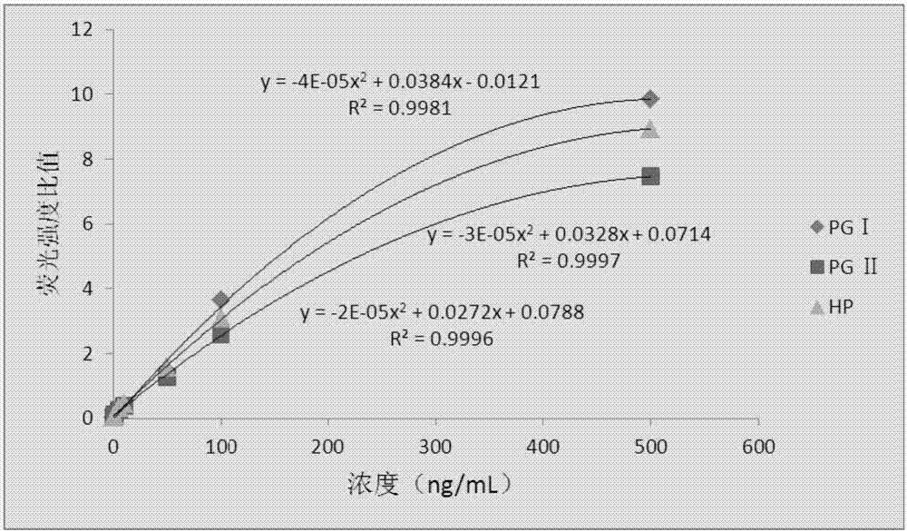
[0070] (1) Detection sensitivity
[0071] Use pepsinogen I, pepsinogen II antigen, and anti-Helicobacter pylori antibody as the samples to be tested to determine the detection of pepsinogen I, pepsinogen II, and anti-Helicobacter pylori antibody lateral flow detection reagent in Example 1. sensitivity.
[0072] Pepsinogen Ⅰ, pepsinogen Ⅱ antigen, and anti-Helicobacter pylori antibody were prepared with 0.02M PBS buffer solution at pH 7.4 containing 5% calf serum to a series of concentrations (0, 0.5, 1, 5, 10, 50, 100, 500 ng / mL), respectively added to the loading end of the detection pepsinogen I, pepsinogen II antigen, and anti-Helicobacter pylori antibody lateral flow detection reagent obtained in Example 1, and detected by fluorescence The instrument detects the fluorescence intensity. Detection steps: return the sample to be tested to room temperature (25°C) before the test, a...
PUM
| Property | Measurement | Unit |
|---|---|---|
| Particle size | aaaaa | aaaaa |
| Sensitivity | aaaaa | aaaaa |
Abstract
Description
Claims
Application Information
 - R&D
- Intellectual Property
- Life Sciences
- Materials
- Tech Scout
- Unparalleled Data Quality
- Higher Quality Content
- 60% Fewer Hallucinations
Browse by: Latest US Patents, China's latest patents, Technical Efficacy Thesaurus, Application Domain, Technology Topic, Popular Technical Reports.
© 2025 PatSnap. All rights reserved.Legal|Privacy policy|Modern Slavery Act Transparency Statement|Sitemap|About US| Contact US: help@patsnap.com