Tumor microvascular imaging instrument and tumor microvascular imaging method

A technology of microvessels and imagers, applied in the field of biomedicine, can solve the problems of tens of microns of capillary pipeline difficulties, biological tissues limit the imaging depth of optical imaging methods, etc.

Pending Publication Date: 2017-12-01
WUHAN UNIV
View PDF6 Cites 13 Cited by
  • Summary
  • Abstract
  • Description
  • Claims
  • Application Information

AI Technical Summary

Problems solved by technology

At present, the clinical imaging methods for blood vessels mainly include ultrasound, CT angiography and nuclear magnetic angiography. However, it is still very difficult for these methods to distinguish capillary channels of tens of microns.
[0005] At present, optical imaging methods that are gradually beginning to b

Method used


Image

  • Tumor microvascular imaging instrument and tumor microvascular imaging method

Examples


Embodiment Construction

[0015] In order to make the objectives, technical solutions and advantages of the present invention clearer, the present invention will be described in further detail below in conjunction with the accompanying drawings and embodiments. It should be understood that the specific embodiments described here are only used to explain the present invention and are not intended to To limit the present invention, in addition, the technical features involved in the various embodiments of the present invention described below can be combined with each other as long as they do not constitute conflicts with each other.

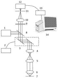
[0016] please see figure 1 , a tumor microvascular imager provided by the present invention includes an infrared confocal imaging part and a control part; the near-infrared confocal imaging part includes a near-infrared laser 1, a dichroic mirror 8, a two-dimensional scanning galvanometer 3, and a scanning lens group , imaging objective lens 6, near-infrared fluorescence f...


PUM


Abstract

The invention discloses a tumor microvascular imaging instrument and a tumor microvascular imaging method. The tumor microvascular imaging instrument comprises an infrared confocal imaging part and a control part, wherein the infrared confocal imaging part comprises a near-infrared laser, a dichroscope, a two-dimensional scanning galvanometer, a scanning lens system, an imaging objective, a near-infrared fluorescence filter, a convergent lens, a pinhole and a detector; the scanning lens system is composed of a field lens and a cylindrical lens; the control part comprises a two-dimensional scanning control module which is used for controlling the two-dimensional scanning galvanometer, a signal amplification and acquisition module and a data processing and image displaying module; near-infrared quantum dots that a fluorescence excitation spectrum ranges from 1000nm to 1350nm and a fluorescence emission spectrum ranges from 1350nm to 1500nm are injected to blood vessels of a to-be-observed living body; and then a tumor part of the living body is observed under the tumor microvascular imaging instrument. According to the tumor microvascular imaging instrument and the tumor microvascular imaging method provided by the invention, complete three-dimensional imaging of tumor microvessels of the living body can be implemented with a high resolution.

Description

technical field [0001] The invention belongs to the technical field of biomedicine, and relates to a tumor microvessel imager and a tumor microvessel imaging method, in particular to an imaging instrument with a working wavelength range of 1300-1500nm and a tumor microvessel imaging method. Background technique [0002] The formation of tumor microvessels has a very important impact on the growth and invasion and metastasis of tumors, especially on the early occurrence of tumors. Studies have shown that the rapid growth of tumors, especially malignant tumors, depends on angiogenesis. Some tumors are rich in microvessels, immature in shape, complex in network structure, and have obvious heterogeneity in composition, shape and spatial distribution, that is, phenotypic heterogeneity of tumor microvessel architecture. This heterogeneity may be derived from the imbalance of various angiogenesis-promoting and inhibitory factors in the regulatory mechanism of tumor angiogenesis. T...

Claims


Application Information

Patent Timeline
IPC IPC(8): A61B5/00
CPCA61B5/0064A61B5/0071A61B5/0073A61B5/0075A61B5/0077
Inventor 黄凯徐海波
Owner WUHAN UNIV
Why Eureka
Try Eureka