Composite stent as well as preparation method and application thereof

A composite stent and composite coating technology, applied in the field of biomedical materials, can solve problems that have not been solved, and achieve the effect of simple structure, good biocompatibility, and increased hydrophilicity

Inactive Publication Date: 2018-03-30
PEKING UNION MEDICAL COLLEGE HOSPITAL CHINESE ACAD OF MEDICAL SCI
View PDF5 Cites 11 Cited by
  • Summary
  • Abstract
  • Description
  • Claims
  • Application Information

AI Technical Summary

Problems solved by technology

However, how to load it onto various 3D printed scaffolds and make it osteogenic and allow cells to interact with it has not been resolved.

Method used


Image

  • Composite stent as well as preparation method and application thereof
  • Composite stent as well as preparation method and application thereof
  • Composite stent as well as preparation method and application thereof

Examples


Embodiment 1

[0058] 1. Preparation of three-dimensional porous metal scaffold

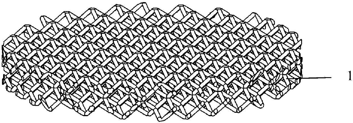
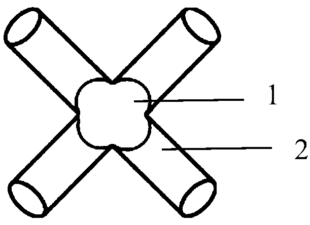
[0059] (1) Import the CT image into the CAD software, and use the CAD software to build a 3D digital model with a rhombic dodecahedron as the basic unit, a hole diameter of 100 μm, and a hole column of 200 μm.

[0060] (2) Input the above preparation parameters into the laser 3D printing equipment (ConceptLaser Germany), the maximum scanning speed is 7m / s, and the construction speed is 1-5cm 3 / h. Set the printing layer thickness, melting speed, scanning direction, laser spot interval, powder spreading speed, melt the powder, build up layer by layer, and then use wire cutting to separate the sample and heat treatment to release stress.

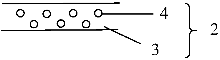
[0061] (3) Soak in 10% dilute hydrochloric acid overnight, and ultrasonically clean to remove residual titanium alloy powder to obtain a three-dimensional porous titanium alloy stent. Its structure is as follows figure 1 Shown.

[0062] 2. Preparation of hyaluronic acid-dopamine

[0063...


PUM

PropertyMeasurementUnit
pore sizeaaaaaaaaaa
thicknessaaaaaaaaaa
thicknessaaaaaaaaaa

Abstract

The invention discloses a composite stent, which comprises a three-dimensional porous stent and a composite coating, wherein the three-dimensional porous stent is of a three-dimensional communicated porous reticular structure; the composite coating main body structure is of a gel layer formed by hyaluronic acid-dopamine; the composite coating also comprises functional nanometer particles; the functional nanometer particles are uniformly distributed in the gel layer; the composite coating is adhered onto the surface of the three-dimensional porous stent. The invention also provides a preparation method of the composite stent. The composite stent can be applied to the preparation of a bone defect restoration material; the requirement of the bone restoration material on the mechanical strength is met; better biocompatibility and hydrophilicity are realized; the material after the coating is favorable for the cell adhesion; the inside functional nanometer particles are favorable for the deviation in directions of ossification, vascularization and the like; the tissue restoration is promoted.

Description

Technical field [0001] The invention relates to the technical field of biomedical materials, in particular to a composite scaffold and a preparation method and application thereof. Background technique [0002] Bone defects are often caused by fractures, bone tumors, bone and joint infections and other diseases. The treatment is very difficult. In severe cases, it can lead to lifelong disability. Due to the limitation of the source of bone grafts, especially for large-scale bone defects and irregular bone defects, the current clinical treatment Upgrading is still a difficult problem. Many patients even have their limbs amputated due to bone defects. The shortcomings of autologous bone transplantation limit its use, including: donor site pain, paresthesia, bleeding, non-union infection and other problems. From 1999 to 2008, the use of autologous bone transplantation in orthopedic surgery dropped from 86% to 10%; the current bone transfer technology can treat bone defects, but th...

Claims


Application Information

Patent Timeline
Patent Type & Authority Applications(China)
IPC IPC(8): A61L27/34A61L27/30A61L27/28A61L27/06A61L27/50A61L27/54A61L27/56B33Y10/00B33Y70/00
CPCA61L27/06A61L27/28A61L27/306A61L27/34A61L27/50A61L27/54A61L27/56B33Y10/00B33Y70/00A61L2300/102A61L2430/02A61L2420/04A61L2420/06A61L2400/12A61L2300/412A61L2300/602C08L5/08
Inventor 于凌佳黄振飞贺宇王海吴志宏马步鹏林进
Owner PEKING UNION MEDICAL COLLEGE HOSPITAL CHINESE ACAD OF MEDICAL SCI
Why Eureka
Try Eureka