Method for preparing skin tissue engineering scaffold based on 3D bio-printing technology and in-vitro cytotoxicity testing method for scaffold

A technology of skin tissue engineering and tissue engineering scaffold, which is applied in the field of 3D printing technology, can solve the problems of fast degradation speed and poor mechanical properties, and achieve the effect of easy control, good mechanical properties and improved mechanical strength

Inactive Publication Date: 2018-06-05
YANGZHOU UNIV
View PDF3 Cites 6 Cited by
  • Summary
  • Abstract
  • Description
  • Claims
  • Application Information

AI Technical Summary

Problems solved by technology

[0004] The research and selection of scaffold materials is an important research content of tissue engineering scaffolds. Gelatin, a natural polymer material, has good biocompatibility, biodegradability and bioactivity, and can better induce self-recovery of injured skin, b

Method used


Image

  • Method for preparing skin tissue engineering scaffold based on 3D bio-printing technology and in-vitro cytotoxicity testing method for scaffold
  • Method for preparing skin tissue engineering scaffold based on 3D bio-printing technology and in-vitro cytotoxicity testing method for scaffold
  • Method for preparing skin tissue engineering scaffold based on 3D bio-printing technology and in-vitro cytotoxicity testing method for scaffold

Examples


Embodiment 1

[0033] (1) Preparation of gelatin (gelatin, GEL) solution: add gelatin to phosphate buffered solution (PBS), and keep stirring until the gelatin is completely dissolved;

[0034] (2) Preparation of cellulose nanofibers (cellulose nanofibers) / gelatin (gelatin, GEL) composite hydrogel: Add a certain amount of CNF solution to the GEL solution, so that the GEL concentration is 3%, and the CNF solid filling amount is 3%. %, blending and stirring for 1 hour at 30°C;

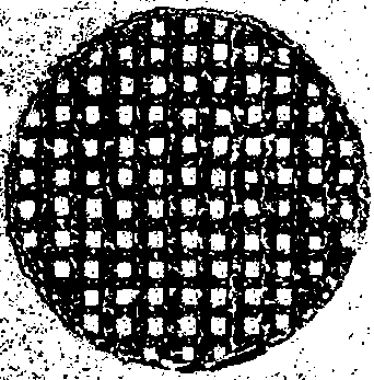
[0035] (3) 3D printing of tissue engineering scaffolds: Put the CNF / GEL composite hydrogel material prepared above into the barrel of a 3D printer, set the barrel temperature to 3°C, platform temperature to 1°C, needle diameter to 150 μm, and extrude The pressure is 0.08MPa, the travel speed of the nozzle is 10mm / S, and the aperture is 100μm to complete the 3D printing of tissue engineering scaffolds;

[0036] (4) Cross-linking of tissue engineering scaffolds: Put the printed scaffolds into the genipin solution with a...

Embodiment 2

[0038] (1) Preparation of gelatin (gelatin, GEL) solution: add gelatin to phosphate buffered solution (PBS), and keep stirring until the gelatin is completely dissolved;

[0039] (2) Preparation of cellulose nanofibers (cellulose nanofibers) / gelatin (gelatin, GEL) composite hydrogel: Add a certain amount of CNF solution to the GEL solution so that the GEL concentration is 5%, and the CNF solid filling amount is 5%. %, blending and stirring for 2 hours at 30°C;

[0040] (3) 3D printing of tissue engineering scaffolds: Put the CNF / GEL composite hydrogel material prepared above into the barrel of a 3D printer, set the barrel temperature to 5°C, platform temperature to 3°C, needle diameter to 150 μm, and extrude The pressure is 0.1MPa, the nozzle walking speed is 10mm / S, and the aperture is 150μm, and the 3D printing of tissue engineering scaffold is completed;

[0041] (4) Cross-linking of tissue engineering scaffolds: Put the printed scaffolds into the genipin solution with a c...

Embodiment 3

[0043](1) Preparation of gelatin (gelatin, GEL) solution: add gelatin to phosphate buffered solution (PBS), and keep stirring until the gelatin is completely dissolved;

[0044] (2) Preparation of cellulose nanofibers (cellulose nanofibers) / gelatin (gelatin, GEL) composite hydrogel: Add a certain amount of CNF solution to the GEL solution so that the GEL concentration is 6%, and the CNF solid filling amount is 8% %, blending and stirring for 3 hours at 35°C;

[0045] (3) 3D printing of tissue engineering scaffolds: put the CNF / GEL composite hydrogel material prepared above into the barrel of a 3D printer, set the barrel temperature to 8°C, platform temperature to 5°C, needle diameter to 220 μm, and extrude The pressure is 0.1MPa, the traveling speed of the nozzle is 15mm / S, and the aperture is 200μm to complete the 3D printing of tissue engineering scaffolds;

[0046] (4) Cross-linking of tissue engineering scaffolds: Put the printed scaffolds into the genipin solution with a...


PUM

PropertyMeasurementUnit
Apertureaaaaaaaaaa
Diameteraaaaaaaaaa
Heightaaaaaaaaaa

Abstract

The invention discloses a method for preparing a skin tissue engineering scaffold based on a 3D bio-printing technology and an in-vitro cytotoxicity testing method for the scaffold. The method comprises the following process steps: preparing high-strength cellulose nanofiber/gelatin complex hydrogel for a printing scaffold, printing a 3D tissue engineering scaffold and cross-linking the scaffold.According to the method disclosed by the invention, the requirements on high porosity and high precision of the tissue engineering scaffold are met by utilizing the 3D bio-printing technology. The CNFserves as a filling material of the GEL and has an effect of improving the mechanical strength of the GEL, and the printed scaffold is soaked in a genipin solution to be cross-linked. The skin tissueengineering scaffold prepared by the method has excellent mechanical property, and does not have any toxic or side effect or immunologic rejection. Meanwhile, the tissue engineering scaffold preparedby using the 3D printing technology has the advantages of being convenient, rapid and easy to control, and can be subjected to personalized customization according to depths, sizes and shapes of wounds of patients.

Description

technical field [0001] The invention relates to a method for preparing a skin tissue engineering scaffold based on 3D bioprinting technology and an in vitro cytotoxicity test method of the scaffold, in particular to a biocompatibility, biodegradability, biological activity and meeting the requirements of tissue engineering scaffolds The preparation of composite materials and the corresponding 3D printing process. Background technique [0002] Tissue defect repair and organ transplantation are one of the greatest breakthroughs in the history of medicine in the 20th century. The emergence of bio-3D printing technology has brought a new dawn to tens of millions of patients who need organ transplantation and tissue repair. [0003] 3D printing, also known as rapid prototyping technology, was born in the late 1980s. This is a manufacturing technique based on the material build-up method, which is considered a major achievement in manufacturing in the past 20 years. It is a sci...

Claims


Application Information

Patent Timeline
IPC IPC(8): A61L27/60A61L27/56A61L27/52A61L27/50A61L27/58A61L27/26B33Y10/00B33Y70/00G01N33/50
CPCA61L27/26A61L27/50A61L27/52A61L27/56A61L27/58A61L27/60B33Y10/00B33Y70/00G01N33/5014C08L89/00C08L1/02
Inventor 周骥平姜亚妮许晓东张琦赵国琦朱兴龙
Owner YANGZHOU UNIV
Why Eureka
Try Eureka