Time resolution fluorescence immunochromatography test strip and kit for jointly detecting CA19-9 and Carcino-Embryonic Antigen (CEA)

A technology of time-resolved fluorescence and immunochromatography test strips, which is applied in the direction of analytical materials, biological testing, measuring devices, etc., can solve problems such as inappropriate diagnosis, and achieve the effects of stable quality, simple equipment, and good detection performance

Inactive Publication Date: 2018-06-08
河南省生物工程技术研究中心
View PDF5 Cites 12 Cited by
  • Summary
  • Abstract
  • Description
  • Claims
  • Application Information

AI Technical Summary

Problems solved by technology

[0005] At present, domestic detection methods for CA19-9 and CEA include radioimmunoassay, chemiluminescence, electrochemiluminescence, enzyme-linked immu

Method used


Image

  • Time resolution fluorescence immunochromatography test strip and kit for jointly detecting CA19-9 and Carcino-Embryonic Antigen (CEA)
  • Time resolution fluorescence immunochromatography test strip and kit for jointly detecting CA19-9 and Carcino-Embryonic Antigen (CEA)
  • Time resolution fluorescence immunochromatography test strip and kit for jointly detecting CA19-9 and Carcino-Embryonic Antigen (CEA)

Examples


Embodiment 1

[0033] Carboxyl group fluorescent microspheres are used as composite raw materials, polystyrene-carboxyeuropium chelate, the solid content is 1%, the particle size is 210nm, the maximum emission wavelength is 613nm, and the maximum excitation wavelength is 365nm.

[0034] Wash the time-resolved fluorescent microspheres twice with activation buffer (0.05M boric acid buffer at pH 8.0), resuspend with activation buffer, add carbodiimide, shake and activate at 30°C for 60min, add 20ul of 10% ethanol After mixing, centrifuge and wash to remove carbodiimide, add labeling buffer (0.05M boric acid buffer at pH 8.0), mix well, then add 80ug CA19-9 monoclonal antibody and 65ug CEA monoclonal antibody, shake at 37°C Mark for 2 hours, and after centrifugation, use reconstitution buffer (0.01MTris-Hcl (pH8.5), 0.1%BSA, 0.1%T-20, 0.1%PVP, 0.1%PEG20000, 3% trehalose, 0.05%NaN 3 ) resuspended, added 50ul of 10% BSA to block for 30min, and obtained time-resolved fluorescent microsphere-labeled...

Embodiment 2

[0036] CA19-9 monoclonal antibody (denoted as S1 (subtype IgG2a, titer 1:128), S2 (subtype IgG3, titer 1:256), S3 (subtype IgG2a, titer 1 :256), S4 (subtype IgG1, titer 1:128)) and CEA monoclonal antibody (denoted as M1 (subtype IgG3, titer 1:128), M2 (subtype IgG2a, potency 1:128) 1:256), M3 (subtype IgG2a, titer 1:128), M4 (subtype IgG2a, titer 1:64)) were provided by Henan Bioengineering Technology Research Center. The method in Example 1 was used to couple with time-resolved fluorescent microspheres to prepare labeled CA19-9 monoclonal antibody complexes (referred to as S1 complex, S2 complex, S3 complex, and S4 complex) and CEA monoclonal antibody complexes. Clone antibody complexes (denoted M1 complexes, M2 complexes, M3 complexes, M4 complexes). The labeled CA19-9 monoclonal antibody complex and CEA monoclonal antibody complex were mixed and sprayed on glass fiber cotton, and the CA19-9 monoclonal antibody and CEA monoclonal antibody were respectively coated on the nit...

Embodiment 3

[0042] Example 3: Preparation of time-resolved fluorescent immunochromatographic test strips for joint detection of CA19-9 and CEA

[0043] (1) Using the method in Example 1, prepare time-resolved fluorescent microsphere-labeled CA19-9 monoclonal antibody S2 complex and CEA monoclonal antibody M1 complex;

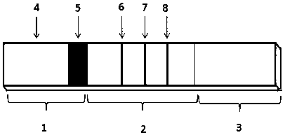
[0044] (2) Antibody coating of detection line and quality control line: Paste the nitrocellulose membrane on the middle of the bottom plate, and overlap with the absorbent paper on the upper end by 1-2mm, and use 0.05M PBS, 1%BSA, 0.05% NaN 3 The CA19-9 monoclonal antibody S4, CEA monoclonal antibody M2 and goat anti-mouse polyclonal antibody diluted in the diluent were coated on the T1 line, T2 line and C line position on the nitrocellulose membrane respectively. Under 40% humidity, dry for 4 hours;

[0045] (3) Spraying in the bonding area: Soak the glass fiber cotton in the treatment solution (0.01MTris-Hcl (pH8.5), 0.1%BSA, 0.1%T-20, 0.1%PVP, 0.1%PEG20000, 3%Trehalose...


PUM

PropertyMeasurementUnit
Particle sizeaaaaaaaaaa

Abstract

The invention relates to a time resolution fluorescence immunochromatography test strip for jointly detecting CA19-9 and a Carcino-Embryonic Antigen (CEA). The test strip comprises a base plate, a nitrocellulose membrane, a glass fiber cotton and water absorption paper, wherein the nitrocellulose membrane is coated with a CA19-9 monoclonal antibody used as a test 1 wire (T1 wire), a CEA monoclonalantibody used as a test 2 wire (T2 wire) and a sheep anti-mouse polyclonal antibody used as a quality control wire (C wire); the glass fiber cotton is divided into a sample application region and a combination region; the combination region is sprayed with a time resolution fluorescence microsphere-labeled CA19-9 monoclonal antibody compound and a CEA monoclonal antibody compound. According to the time resolution fluorescence immunochromatography test strip and the kit for jointly detecting the CA19-9 and the CEA, test for the concentrations of the CA19-9 and the CEA can be completed within arelatively short time; a test result is high in accuracy and high in sensitivity; a requirement for quick test beside a clinical bed and requirements of a primary community hospital can be met.

Description

technical field [0001] The invention belongs to the technical field of diagnostic kits, and specifically relates to a time-resolved fluorescent immunochromatographic test strip for joint detection of tumor marker sugar chain antigen 19-9 and carcinoembryonic antigen, a kit containing the test strip, and Their use in the diagnosis of pancreatic cancer. Background technique [0002] Carbohydrate antigen 19-9 (CA19-9) is a specific glycoprotein produced by digestive system tumor tissue, also known as sialylated Lewis a antigen. CA19-9 is a digestive system-related antigen, mainly expressed in pancreatic epithelial cells, and its content in normal human serum is less than 37 U / ml. Studies have shown that CA19-9 is abnormally expressed in patients with malignant tumors of the digestive system. Among them, there is a high positive rate in pancreatic cancer, biliary tract cancer, gastric cancer, colon cancer, esophageal cancer, and liver cancer, while CA19-9 has a high positive r...

Claims


Application Information

Patent Timeline
IPC IPC(8): G01N33/533G01N33/558G01N33/577G01N33/574G01N33/68
Inventor 王云龙米亚双李玉林王继创程蕾
Owner 河南省生物工程技术研究中心
Why Eureka
Try Eureka