
Virus isolation method for low-content sample of porcine epidemic diarrhea virus (PEDV)

A porcine epidemic diarrhea and virus technology, applied in the direction of virus, virus/phage, anti-viral immunoglobulin, etc., can solve the problems of difficulty in obtaining success, difficulty in isolating a single pathogen, and achieve the effect of effective separation

Active Publication Date: 2018-09-04
LANZHOU INST OF VETERINARY SCI CHINESE ACAD OF AGRI SCI
View PDF5 Cites 3 Cited by
  • Summary
  • Abstract
  • Description
  • Claims
  • Application Information

AI Technical Summary

Problems solved by technology

However, the current clinical pathogenic infection is complex and diverse, and clinical cases of single pathogenic infection have been rare. In this case, it is very difficult to isolate a single pathogenic agent using conventional methods
At the same time, for some samples with low pathogenic content, it is difficult to successfully separate them using conventional methods

Method used


Image

  • Virus isolation method for low-content sample of porcine epidemic diarrhea virus (PEDV)
  • Virus isolation method for low-content sample of porcine epidemic diarrhea virus (PEDV)
  • Virus isolation method for low-content sample of porcine epidemic diarrhea virus (PEDV)

Examples


Embodiment 1

[0039] Example 1 Preparation of PEDV-specific immune nano-magnetic beads

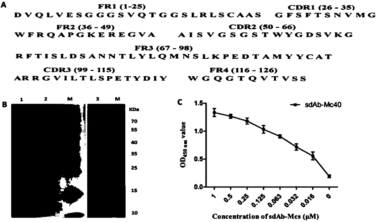
[0040] 1.1 Screening and expression of PEDV membrane protein (M) specific sdAb

[0041] Using phage display technology, panned the M protein immune Bactrian camel library, obtained the phage that specifically binds to the M protein, performed sequencing analysis, obtained the sdAb gene sequence, and its nucleotide sequence is shown in SEQ ID NO.1, and performed sequence analysis Then it is cloned into PET-32a expression plasmid, expressed with Escherichia coli (E.coli), and utilizes Ni purification resin to carry out target protein purification; By enzyme-linked immunosorbent assay (ELISA), analyze sdAb-Mc40 and M protein binding activity.

[0042] Phage sequencing analysis of M protein binding to obtain specific single domain antibody sdAb-Mc40( figure 1 A); After the 32a vector is connected, it is transformed into Escherichia coli to obtain an expressed protein of consistent size, and for soluble ex...

Embodiment 2

[0058] The virus isolation method of embodiment 2 PEDV low content sample

[0059] 1 Clinical sample processing

[0060] For suspected PEDV-infected pig feces and secretions and other cotton swab samples, put them in a 2mL centrifuge tube, add 1mL sterile PBS (pH 7.4) and place at room temperature for 2h, centrifuge at 2000rpm for 5min, and take the supernatant; suspected PEDV-infected dead pig tissue samples, Use sterile PBS (pH 7.4) tissue homogenate (homogenizer or quartz sand grinding), place at room temperature for 4h or overnight at 4°C, centrifuge at 10000rpm for 10min, and take the supernatant.

[0061] 2. Immunization of nano-magnetic beads to capture PEDV in samples

[0062] Aseptically take 20 μL of 1 mg / mL immune nano-magnetic beads, add to a 2 mL centrifuge tube, wash 3 times with sterile PBS (pH 7.4), and collect the immune nano-magnetic beads on a magnetic stand. Take 1mL of the supernatant of the processed sample, add it to the centrifuge tube containing the ...

Embodiment 3

[0065] The virus isolation of embodiment 3 field PEDV infection samples

[0066] After the fecal samples of clinically suspected infection were processed, they were enriched with immune nano-magnetic beads, detected by RT-PCR, and then the captured virus was inoculated into vero cells, and CPE was observed and passaged. At the same time, it was verified by immunofluorescence and cell ultrathin section transmission electron microscopy, and the S gene was cloned by PCR for sequence analysis. Results The suspected PEDV-infected piglet feces samples were processed directly by RT-PCR, and no obvious bands were seen after electrophoresis, but after incubation with different amounts of samples and magnetic beads, RT-PCR detection showed the expected small bands ( Figure 5 B). 24h after inoculating vero cells with the magnetic beads incubated with the sample, obvious CPE appeared ( Figure 5 A); After the virus was passaged, it could be observed that there was obvious fluorescence ...


PUM


Abstract

The invention discloses a virus isolation method for a low-content sample of porcine epidemic diarrhea viruses (PEDV), and further discloses an immune nano-magnetic bead for enrichment and isolation of the PEDV. The immune nano-magnetic bead has a magnetic Fe2O3 core and a surface coupled with PEDV membrane protein-specific single-domain antibodies. Traditional methods for isolating viruses from multiple infected and low PEDV-content samples require viral purification and long-term blind transmission, the separation efficiency is low, and the research work of the PEDV is limited. The virus isolation method, provided by the invention, utilizes nano-magnetic beads and PEDV-specific single-domain antibodies to prepare the immune nano-magnetic beads, and can effectively capture and enrich thePEDV in various samples such as feces and tissues. After the samples are processed, the immune nano-magnetic beads are utilized for virus capture and enrichment, the immune nano-magnetic beads combined with the virus are directly inoculated into Vero cells, and the virus isolation can be achieved. The immune nano-magnetic beads and the PEDV isolation method prepared by the invention are suitable for rapid and effective isolation of the PEDV in clinically complex and low virus-content samples.

Description

technical field [0001] The invention relates to an immune nanometer magnetic bead used for isolating porcine epidemic diarrhea virus (PEDV), and also relates to a method for efficiently separating the virus from clinical samples with low PEDV content. The invention belongs to the field of biotechnology. Background technique [0002] Porcine epidemic diarrhea virus (PEDV) belongs to the Nidovirales order, Coronaviridae family, and a member of the Coronavirus genus. It is the pathogen that causes porcine epidemic diarrhea (PED). PED is a highly contagious intestinal infectious disease mainly characterized by diarrhea, vomiting and dehydration in infected pigs. Pigs of all ages can be infected, especially for suckling piglets with a high mortality rate. The incidence of porcine epidemic diarrhea spreads all over the world, which brings great harm to the pig industry. The rapid isolation and identification of viruses in clinical PED pig samples is a necessary link in the study...

Claims


Application Information

Patent Timeline
IPC IPC(8): C12N7/00C12N7/02C12N15/11C07K16/10H01F1/00H01F41/00
CPCC07K16/10C12N7/00C12N2770/20051H01F1/0054H01F41/00
Inventor 杨顺利尹双辉蔡建平李丽尚佑军刘湘涛
Owner LANZHOU INST OF VETERINARY SCI CHINESE ACAD OF AGRI SCI
Learn More