Tissue repair stent and preparation method and application thereof

A technology for tissue repair and fiber filaments, applied in tissue regeneration, medical science, prostheses, etc., can solve problems such as fibrosis, rejection, and insufficient donors, and achieve tissue regeneration with low possibility and shape easy effect

Active Publication Date: 2018-11-06
FOURTH MILITARY MEDICAL UNIVERSITY +1
View PDF16 Cites 2 Cited by
  • Summary
  • Abstract
  • Description
  • Claims
  • Application Information

AI Technical Summary

Problems solved by technology

[0008] In view of the technical problems existing in the tissue repair process in the prior art, for hard tissue repair, for example: biocompatibility is not superior, bone cell induction performance is insufficient, the scaffold does not match the shape of the defect, etc.; for cartilage repair For example, the shape of the scaffold cannot meet the defect requirements, the mechanical properties of the scaffold do not match those of autologous cartilage, the ability to move cannot be restored as soon as possible, and tissues similar to hyaline cartilage cannot be formed after long-term use; technical problems of fibrosis are prone to occur; there is no long-term natural-like Biological properties of cartilage tissue, joint degeneration will occur within a period of time; donor shortage; poor interface bonding will cause rejection, etc.

Method used


Image

  • Tissue repair stent and preparation method and application thereof
  • Tissue repair stent and preparation method and application thereof
  • Tissue repair stent and preparation method and application thereof

Examples


no. 1 approach

[0044]The first embodiment of the present invention provides a tissue repair scaffold. In the present invention, the tissue repair scaffold may include hard tissue repair scaffolds such as bone repair scaffolds, and may also be cartilage tissue repair scaffolds, bone-cartilage integrated repair scaffolds, and the like. Among them, the bone-cartilage integrated repair scaffold can take care of cartilage repair and subchondral bone repair in full-thickness cartilage defects.

[0045] The tissue repair scaffold of the present invention includes fiber bundles, and the fiber bundles include one or more than two fiber filaments; at least one of the fiber filaments has functional powder on the surface and / or inside. In the present invention, the compression modulus of the tissue repair scaffold can be adjusted according to the defect site of the tissue repair scaffold. Preferably, the compression modulus of the tissue repair scaffold is in the range of 0.1 MPa to 15 MPa, may be in t...

no. 2 approach

[0073] The second embodiment of the present invention provides a method for preparing a tissue repair scaffold, including the step of preparing the tissue repair scaffold by using 3D printing technology; the tissue repair scaffold is the tissue repair scaffold in the first embodiment. The tissue repair scaffold of the present invention adopts the preparation method of precipitation molding, and the dosage ratio between the biodegradable synthetic polymer compound and the functional powder constituting the tissue repair scaffold can be adjusted accordingly according to the needs of the defect site. The parameters such as the average diameter of the tissue repair scaffold, the pore diameter and porosity of the tissue repair scaffold are conducive to meeting the mechanical performance requirements of different defect sites for the tissue repair scaffold, and the biological activity of each component in the tissue repair scaffold is retained during the printing process. Specificall...

no. 3 approach

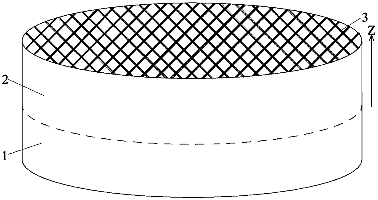
[0086] The third embodiment of the present invention provides a specific tissue repair scaffold prepared in the first embodiment or a specific tissue repair scaffold prepared in the second embodiment, that is, a bone-cartilage integrated repair scaffold. The bone-cartilage integrated repair bracket includes: a bone repair layer 1 and a cartilage repair layer 2 connected. In the present invention, the connected bone repair layer 1 and cartilage repair layer 2 may be in direct contact, or may be connected through other feasible active layers.

[0087] like figure 1 As shown, the bone repair layer 1 and the cartilage repair layer 2 contain fiber bundles 3, and the fiber bundles 3 include one or more than two fiber filaments. The fiber filaments are the fiber filaments in the first embodiment. like image 3 As shown, it can be seen from the electron microscope images that at least one of the fiber filaments has functional powder on the surface and / or inside, preferably, the pa...


PUM

PropertyMeasurementUnit
particle sizeaaaaaaaaaa
pore sizeaaaaaaaaaa

Abstract

The invention provides a tissue repair stent and a preparation method and an application thereof. The tissue repair stent comprises: a fiber bundle comprising one or more filaments, the surface and / orinside of at least one filament is provided with functional powder; the filament material further comprises a biodegradable synthetic high molecular compound. The active ingredient in the functionalpowder of the tissue repair stent can be precipitated in the living body, which is targeted to stimulate the defect locally, and promote tissue regeneration; further, the tissue repair stent is provided with a suitable compressive modulus and the possibility of brittle fracture is small. The preparation method of the tissue repairing stent can adjust the average diameter of the single fiber filament and the pore diameter and porosity of the tissue repairing stent according to the needs of the defect site, thus, the mechanical properties of tissue repair stent at different defect sites can be satisfied, and the biological activities of the components in the stent can be retained in the printing process.

Description

technical field [0001] The invention relates to a tissue repair bracket and its preparation method and application, belonging to the field of medical implant materials. Background technique [0002] Bone defect is a common disease in orthopedic clinic. In clinical operations, bone repair materials are usually used to fill the defect to promote the regeneration of autologous bone tissue, thereby achieving the purpose of bone repair. However, the current bone repair materials have problems such as poor biocompatibility, insufficient bone cell induction performance, and inconsistent shape of actual bone defects in clinical practice, which cannot achieve good bone repair. [0003] In the prior art, the purpose of bone repair is also achieved through bone grafting. Although bone grafting can fill the bone cavity and accelerate the healing of bone defects, autologous bone grafting has been widely recognized by clinicians as the "gold standard" of bone filling technology. There a...

Claims


Application Information

Patent Timeline
Patent Type & Authority Applications(China)
IPC IPC(8): A61L27/58A61L27/54A61L27/50A61L27/18
CPCA61L27/18A61L27/50A61L27/54A61L27/58A61L2300/252A61L2300/414A61L2430/02A61L2430/06C08L67/04
Inventor 陈红庆
Owner FOURTH MILITARY MEDICAL UNIVERSITY
Why Eureka
Try Eureka