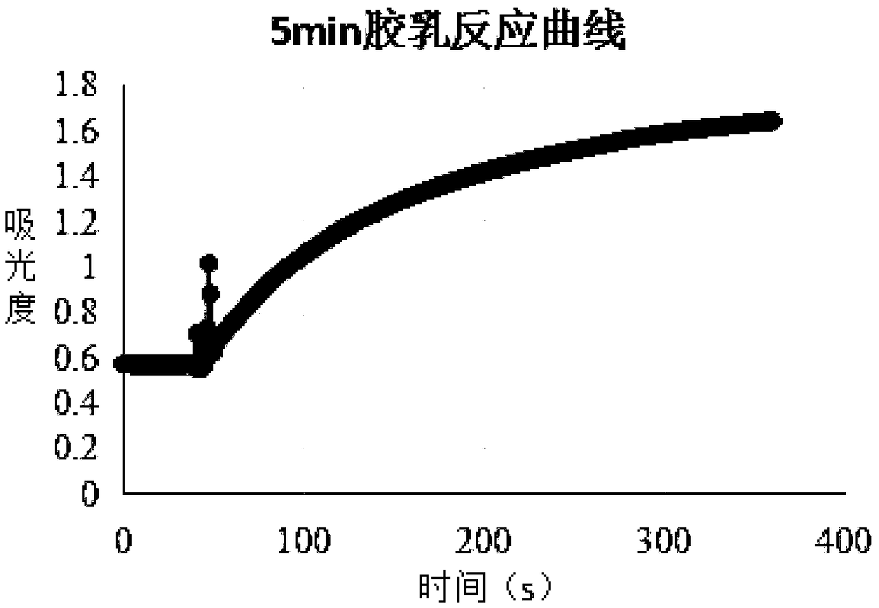
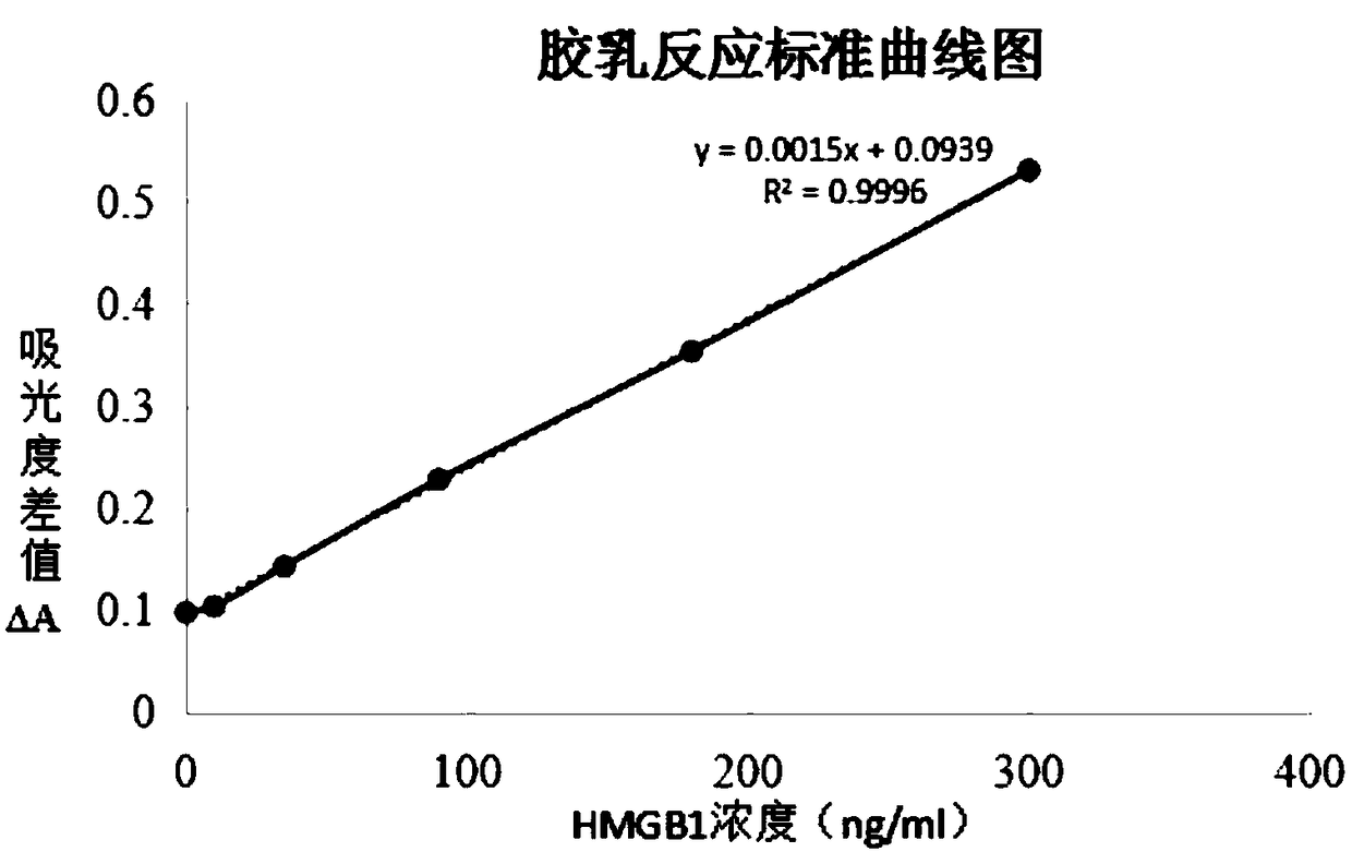
HMGB1 detection kit and preparation method thereof
A technology for detection kits and reagents, applied in the field of medical detection, can solve the problems of uneven quality of detection kits, expensive imported reagents, complicated operation, etc., to speed up time and adsorption equilibrium stability, save reagent costs and sensitivity. high effect
- Summary
- Abstract
- Description
- Claims
- Application Information
AI Technical Summary
Problems solved by technology
Method used
Image
Examples
Embodiment 1
[0058] Example 1 Preparation of HMGB1 antigen
[0059] Recombinant plasmids were constructed, positive recombinant plasmids were screened, and bacteria highly expressing His-SBP-HMGB1 fusion protein were cultivated. Under the conditions of 6000×g and 4°C, centrifuge the culture solution containing bacteria for 30 minutes, collect the bacteria and store them at -20°C for later use; take an appropriate amount of bacteria and resuspend them in the bacteriostasis buffer (containing 50mmol / L Tris-HCL, 250mmol / L Tris-HCL, 250mmol / L L NaCl and 10mmol / L imidazole, pH value is 8.0), sonicate in ice bath, centrifuge at 10000×g, 4℃ for 20min, and collect the supernatant for later use; The non-specific binding protein was washed with bacteria buffer, and then eluted with elution buffer (containing 50mmol / L Tris-HCl, 250mmol / L NaCl and 300mmol / L imidazole, pH value 8.0), and the target protein—HMGB1 was collected, Reserved as HMGB1 antigen. The collected protein concentration was tested ...
Embodiment 2
[0060] Example 2 Preparation of HMGB1 polyclonal antibody
[0061] Dilute the HMGB1 antigen with physiological saline to a concentration of 0.5mg / ml-2mg / ml, and mix it with Freund's adjuvant at a ratio of 1:1. The mixture was injected subcutaneously at multiple points and intramuscularly in the hind legs, and the dose for the first injection was 0.5 mg to 1 mg. Serum was collected every 1 to 2 weeks, and the titer of the serum was determined by agar diffusion test. If the titer did not reach the expected titer, a booster immunization was carried out, and the additional injection dose was 1 / 2 of the first injection. Generally, 2 to 4 booster immunizations were required. When the serum titer reaches the requirement, the rabbit serum is collected, and the collected serum is stored at -20°C for later use.
[0062] Add 100 mg of HMGB1 to 3 g of cyanogen bromide-activated agarose medium (CNBr-activated Sepharose 4B), prepare an HMGB1 affinity chromatography column with a volume of ...
Embodiment 3
[0063] Example 3 Labeling latex particles
[0064] a. Prepare latex solution
[0065] Prepare 1 mL latex solution (22 μL latex mother solution + 978 μL 50 mM MES buffer solution) with a final concentration of 0.11 v / v % with the latex mother solution (5 v / v%, 20 nm ~ 50 nm particle size), the pH value is 6.0.
[0066] b. Activate latex group
[0067] Add three solutions in order to 1mL latex solution: 4.5μL surfactant (5% stock solution prepared with 50mM MES, pH 6.0 solution or pure water); 4.5μL N-HS solution (concentration: 10mg / mL) ; 4.5 μL EDC solution (10 mg / ml concentration). After the solution was added, the latex solution was allowed to stand at 37°C for 1 hour.
[0068] c. Antibody Conjugation Labeling
[0069] 100 μg of HMGB1 antibody (20 μL of 5 mg / mL antibody stock solution) was added, reacted at 37° C. for 2 h, and stirred slowly.
[0070] d. Closed latex group
[0071] 100 μL of blocking solution (1M Tris-HCl, pH 8.0) was added and allowed to stand at 37° ...
PUM
| Property | Measurement | Unit |
|---|---|---|
| diameter | aaaaa | aaaaa |
Abstract
Description
Claims
Application Information
 - R&D
- Intellectual Property
- Life Sciences
- Materials
- Tech Scout
- Unparalleled Data Quality
- Higher Quality Content
- 60% Fewer Hallucinations
Browse by: Latest US Patents, China's latest patents, Technical Efficacy Thesaurus, Application Domain, Technology Topic, Popular Technical Reports.
© 2025 PatSnap. All rights reserved.Legal|Privacy policy|Modern Slavery Act Transparency Statement|Sitemap|About US| Contact US: help@patsnap.com


