Enveloped virus resistant to complement inactivation for the treatment of cancer

A technology for transmembrane domains and proteins, applied in the field of enveloped viruses that are resistant to complement inactivation for the treatment of cancer, can solve problems such as NDV particle destruction

Pending Publication Date: 2019-03-01
WELLSTAT IMMUNOTHERAPEUTICS LLC
View PDF3 Cites 1 Cited by
  • Summary
  • Abstract
  • Description
  • Claims
  • Application Information

AI Technical Summary

Problems solved by technology

Despite the promising initial clinical results, a drawback of NDV as a cancer therapeutic is that once the virus enters th...

Method used


Image

  • Enveloped virus resistant to complement inactivation for the treatment of cancer
  • Enveloped virus resistant to complement inactivation for the treatment of cancer
  • Enveloped virus resistant to complement inactivation for the treatment of cancer

Examples


Embodiment 1

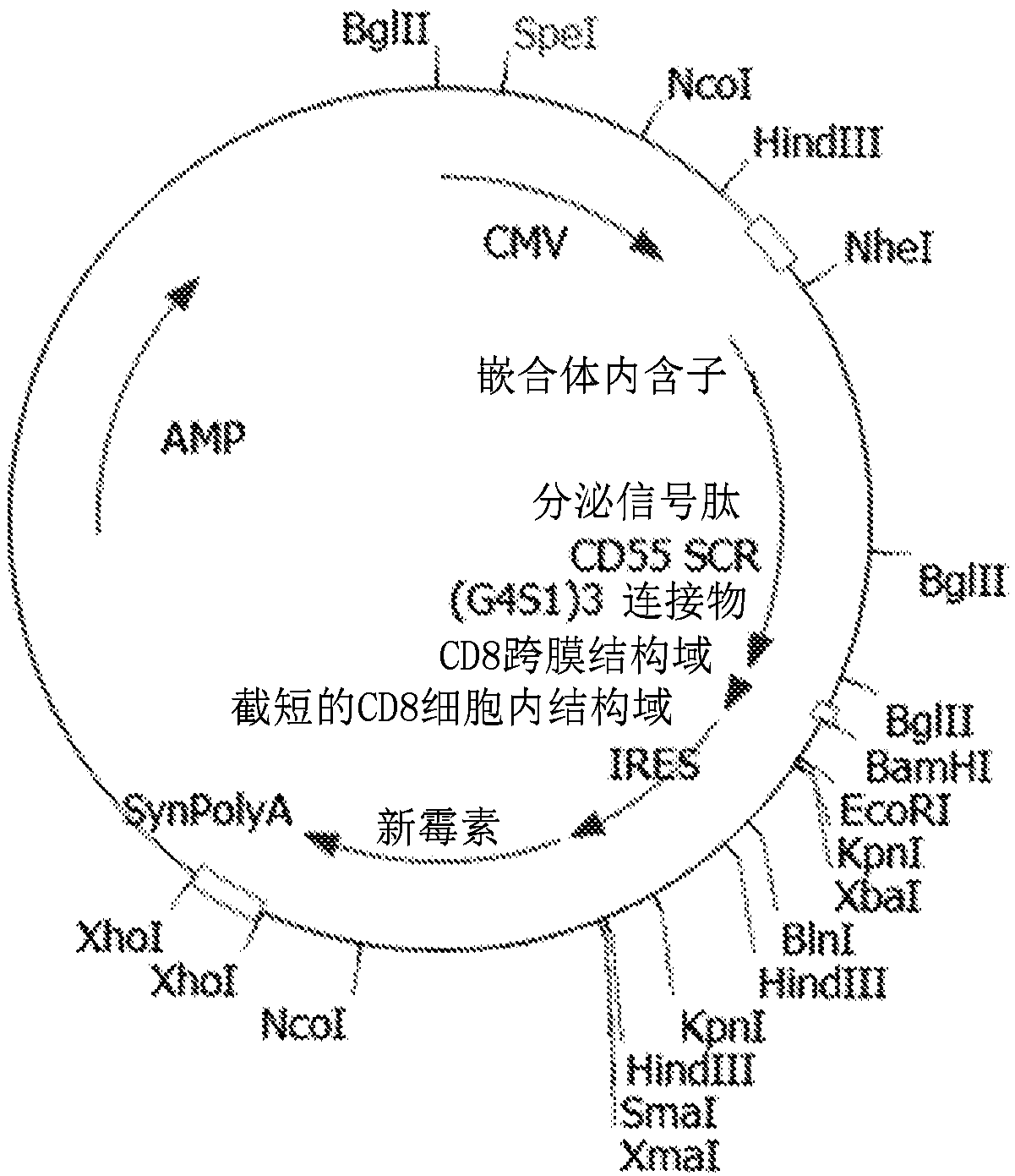
[0035] A modified version of recombinant CD55 was generated with the four short consensus repeats (SCRs) of CD55 downstream of the secretion signal peptide, followed by a flexible linker (3 × G4S1) and the CD8 transmembrane domain and a truncated CD8 intracellular domain ( figure 1 ). The coding sequences were cloned into mammalian expression constructs with CMV promoters (synthetic introns driving expression of recombinant proteins). The expression cassette also contains a drug selectable marker: neomycin phosphotransferase located downstream of the IRES. At the end of the gene expression cassette is a synthetic polyadenylation signal. SEQ ID NO: 1 is the nucleotide sequence of the mammalian cell expression construct. SEQ ID NO: 2 is the amino acid sequence of the expressed protein. When expressed on the cell surface of chicken embryonic fibroblast DF1 or incorporated into the viral membrane, the signal peptide is cleaved, resulting in a mature recombinant fusion protein ...

Embodiment 2

[0037] Mammalian expression constructs were transfected into chicken embryonic fibroblast DF1 cells by PEI 25K (polyethyleneimine, linear 25 kDa, Polysciences, cat# 23966) mediated transfection. 72 hours after transfection, at 300 μg / mLG418 ( aminoglycoside antibiotics) to generate a stable cell line constitutively expressing SEQ ID NO:2. This stable cell line constitutively expressed SEQ ID NO: 3 on its cell surface as detected with a monoclonal antibody specific for mature human CD55 (R&D Systems, catalog # MAB20091). Such as image 3 As shown, recombinant fusion proteins were expressed on DF1 cells stably transfected with constructs encoding recombinant fusion proteins as analyzed by flow cytometry ( image 3 , histogram on the right). Naive DF1 cells served as negative controls ( image 3 , histogram on the left).

Embodiment 3

[0039] A stable cell line expressing SEQ ID NO: 3 on the cell surface was infected with wild-type NDV produced from embryonated chicken eggs. Virus was then titrated on the human tumor cell line HT1080. Equal amounts of virus (measured by PFU) were incubated with 40% normal human serum (NHS) and 40% heat-inactivated normal human serum (iNHS), respectively. Virus surviving incubation with human serum was then scored by plaque assay on HT1080 cells. The ratio of virus recovered after incubation with NHS and with iNHS was calculated. As shown in Table 1, the recovery of virus produced in embryonated eggs was 0.5%, indicating that the vast majority of NDV particles produced by eggs were inactivated, most likely by the human alternative complement pathway. Likewise, the recovery of virus produced by parental chicken embryonic fibroblast DF1 cells was 0.5%. Surprisingly, the recovery rate of virus produced from bulk non-clonal DF1 cells (bulk non-clonal DF1 cells) stably expressi...


PUM


Abstract

A recombinant fusion protein is disclosed. The fusion protein comprises: (a) a CD55 peptide sequence, (b) a linker sequence C-terminal to the CD55 sequence, (c) a transmembrane domain C-terminal to the linker sequence, and (d) an intracellular domain C-terminal to the transmembrane domain. The fusion protein does not contain a GPI anchor. The fusion protein can be expressed with an N-terminal secretory signal peptide, which is cleaved to yield the mature protein on the surface of a cell line or an enveloped virus. An oncolytic virus expressing the fusion protein is resistant to complement inactivation and can be used to treat cancer.

Description

[0001] Sequence Listing Index [0002] The Sequence Listing, file number 21003-PCT, filed electronically as a text file in Annex C / ST.25 is part of this disclosure. Background technique [0003] Oncolytic viruses have been tested as cancer therapeutics by infecting and destroying tumor cells. These oncolytic viruses include Newcastle disease virus, adenovirus, Sindbis virus, vaccinia virus, and herpes virus, among others. Newcastle disease virus (NDV) has shown great potential to shrink tumors in cancer patients due to its unique property of preferentially replicating in tumor cells and lysing tumor cells, presumably due to factors that most tumor cells have a defective interferon pathway (Pecora et al., 2002; Laurie et al., 2006; Lorence et al., 2007). Despite the promising initial clinical results, a drawback of NDV as a cancer therapeutic is that once the virus enters a patient, most NDV particles are inevitably destroyed by the patient's innate immune system - the altern...

Claims


Application Information

Patent Timeline
IPC IPC(8): A61K38/17A61K38/00C07K14/00
CPCA61K38/00A61K35/768C07K19/00A61P35/00C12N15/625C12N2760/18132C12N2760/18152C07K14/70517C07K2319/03C07K2319/036C07K14/70596A61K35/76C12N5/16C12N7/00
Inventor 罗天赐R·莫丽娜G·卡斯蒂尔
Owner WELLSTAT IMMUNOTHERAPEUTICS LLC
Why Eureka
Try Eureka