Application of 18F-SFB-CML and method for detecting atherosclerosis

A technique for atherosclerosis and purpose, applied in the field of nuclear medicine, can solve the problems of ineffective monitoring of plaque composition and vulnerability, poor specificity and sensitivity of F-FDG, limited temporal resolution and spatial resolution, etc. Achieve the effect of being conducive to rapid imaging analysis, not easy to degrade, and easy to high-resolution imaging

Active Publication Date: 2019-03-12
AFFILIATED HOSPITAL OF JIANGSU UNIV
View PDF7 Cites 1 Cited by
  • Summary
  • Abstract
  • Description
  • Claims
  • Application Information

AI Technical Summary

Problems solved by technology

At present, traditional imaging examinations cannot effectively monitor plaque composition and vulnerability, while molecular imaging can accurately determine plaques and has the potential to identify vulnerable and high-risk plaques
[0003] So far, the commonly used methods include (1) nuclear medicine imaging: it is a relatively mature method for molecular imaging of AS plaques. Imaging instruments include positron emission computed tomography (PET) and single photon emission computed tomography (SPECT), but the poor spatial resolution limits the application of this method
(2) CT imaging: However, the temporal resolution and spatial resolution of CT imaging are limited, and the reliability of plaque detection is limited
(3) Magnetic resonance imaging (MR): MR has high spatial resolution and can display fine structures within plaques. The application of targeted contrast agents further improves the early identification of vulnerable plaques. However, MR sequences It is complex, with many influencing factors, and needs to be further explored in large-scale, standardized experiments and clinical studies
(4) Ultrasound examination: Although ultrasound can display lesions dynamically in real time, it is not as good as nuclear medicine, CT and MR in displaying large-scale and multiple vascular lesions
(5) Optical imaging: Optical coherence tomography (OCT) technology has a high spatial resolution and can quantify the components within the plaque, which is of great value in identifying and evaluating the microstructure of vulnerable plaques. However, Limited penetration depth is the main obstacle restricting the application and development of OCT
However using 18 F-FDG diagnostic methods for imaging atherosclerosis have several problems, for example, 18 F-FDG has poor specificity and sensitivity, low imaging speed and enrichment specificity

Method used


Image

  • Application of 18F-SFB-CML and method for detecting atherosclerosis
  • Application of 18F-SFB-CML and method for detecting atherosclerosis
  • Application of 18F-SFB-CML and method for detecting atherosclerosis

Examples


Embodiment 1

[0039] Example 1: 19 Synthesis and quality control of F-SFB-CML standard reference substance

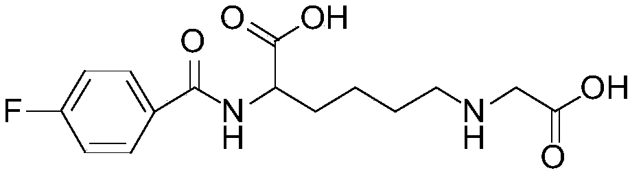
[0040] (1) 19 Synthetic steps of F-SFB-CML standard reference substance

[0041] Take 25 μL of carbonic acid buffer solution (pH=9) of 1 mg / mL CML (carboxymethyllysine complex) dissolved in advance to the reaction tube, and then add [ 19 F]-SFB (N-succinimide-4-fluorobenzoate) solution (1mg / mL) 25μL, then place the reaction tube in an oil bath, react at 65°C for 1h, and use a thin-layer chromatography plate during the reaction After monitoring the reaction, take out the reaction solution and add it to semi-preparative high-performance liquid chromatography (HPLC) for separation and purification, and collect the target product 19 F-SFB-CML. The structure was confirmed by mass spectrometry and NMR, and its chemical purity was detected by analytical high performance liquid chromatography (HPLC).

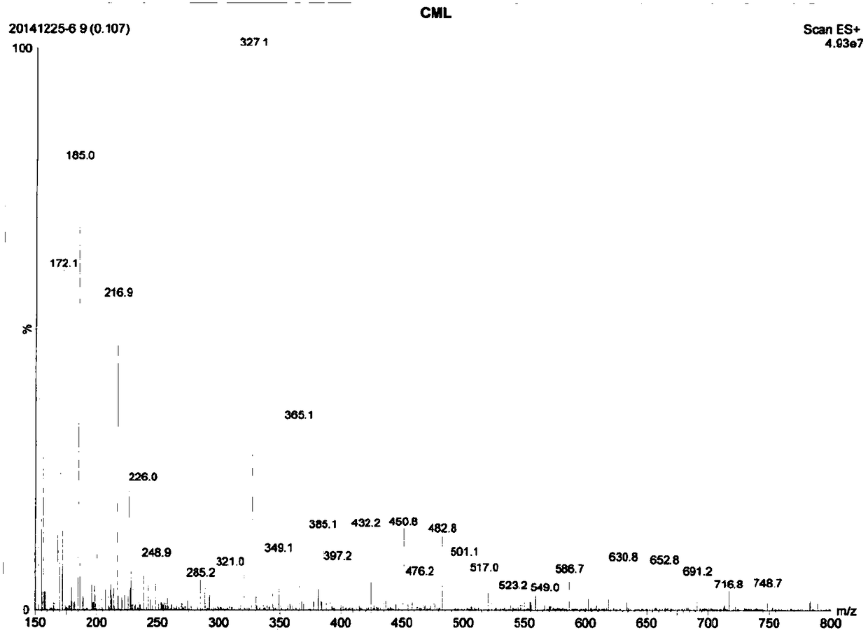
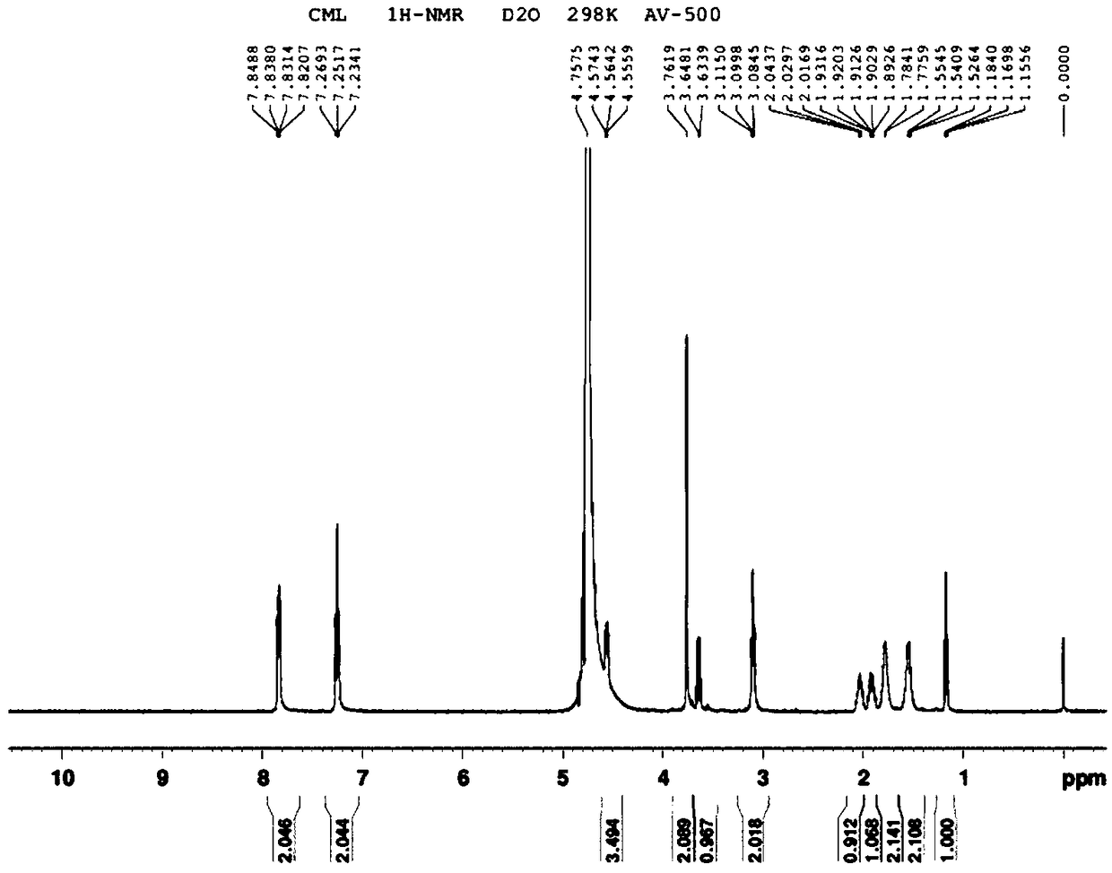
[0042] (2) 19 NMR and MS structure confirmation of F-SFB-CML standard

[0043] obta...

Embodiment 2

[0046] Example 2: 18 F-SFB-CML labeling and isolation and purification

[0047] (1) 18 Radiolabeled synthetic steps of F-SFB-CML

[0048] Produced by bombarding heavy oxygen water with an accelerator 18 F ions first pass through the QMA column (use 10mL 0.5M NaHCO 3 Rinse, and then rinse with 20mL of sterile water for injection) to capture, after enrichment, use K222 / K 2 CO 3 Eluted to reaction tube No. 1, dried to remove water and cooled to room temperature, then added a certain amount of anhydrous acetonitrile to dry and remove water again, and cooled to room temperature.

[0049] Add SFB (10mg SFB, dissolved in 1mL acetonitrile) to react at 90°C for 7min, and cool to room temperature after the reaction. Then 6 mL of 0.1 M HCl was added, and stirred for 1 min. The reaction solution was passed through a C18 column (washed with a mixture of 7.5 mL of 0.1 M HCl and 2.5 mL of acetonitrile before use) to a waste bottle. Add 3 mL of acetonitrile to flow through the C18 colu...

Embodiment 3

[0071] Example 3: ApoE - / - mouse micro PET scan

[0072] (1) Construction of animal model of diabetic atherosclerosis

[0073] Male apoE used in this experiment - / -The mice were fed in the SPF-grade mouse room of the Experimental Animal Center of Jiangsu University. Feeding conditions: temperature 22±2°C, humidity 40-60%, 12-hour cycle lighting, normal diet, free intake of food and water. All items entering the SPF room must be sterilized by high temperature and high pressure, and strict aseptic operation is implemented. At the age of 6 weeks, mice in the experimental group were given intraperitoneal injection of streptozotocin (STZ, dissolved in 0.05 mol / L citrate buffer at pH 4.5) 40 mg / kg / day for 5 consecutive days. After 2 weeks, the mice with blood glucose level > 300 mg / dL were included in the research object of this example, and changed from normal diet to semi-synthetic high-fat diet (high-fat diet, HFD) (21% fat, 0.15% cholesterol, other ingredients same diet), f...


PUM


Abstract

The invention relates to the technical field of nuclear medicine, and relates to application of 18F-SFB-CML and a method for detecting atherosclerosis. Firstly, the invention provides the applicationof 18F-SFB-CML in preparing a product for detecting atherosclerotic diseases; the method for detecting atherosclerosis is further provided; the method is to use 18F-SFB-CML as a photographic developerto obtain the location, size, and vascular stenosis degree information of an atherosclerotic plaque by molecular imaging scanning. The 18F-SFB-CML as the photographic developer can accurately reflectthe size of the plaque and the vascular stenosis degree, and can effectively trace the atherosclerotic plaque.

Description

technical field [0001] The invention relates to the technical field of nuclear medicine, and relates to a 18 Use of F-SFB-CML and method for detecting atherosclerosis. Background technique [0002] Atherosclerosis (AS) is caused by multiple factors, and its pathogenesis is complex. At present, traditional imaging examinations cannot effectively monitor plaque composition and vulnerability, while molecular imaging can accurately determine plaques and has the potential to identify vulnerable and high-risk plaques. [0003] So far, the commonly used methods include (1) nuclear medicine imaging: it is a relatively mature method for molecular imaging of AS plaques. Imaging instruments include positron emission computed tomography (PET) and single photon emission computed tomography (SPECT), but the poor spatial resolution limits the application of this method. (2) CT imaging: However, the temporal resolution and spatial resolution of CT imaging are limited, and the reliability...

Claims


Application Information

Patent Timeline
Patent Type & Authority Applications(China)
IPC IPC(8): A61K51/04A61B6/03A61K101/02
CPCA61B6/03A61B6/481A61B6/482A61B6/504A61K51/0402
Inventor 王中群李丽华严金川邵晨景乐乐张莉莉孙振
Owner AFFILIATED HOSPITAL OF JIANGSU UNIV
Why Eureka
Try Eureka