Fluorescence probe used for pulmonary fibrosis dyeing

A pulmonary fibrosis and fluorescent probe technology, applied in the field of fluorescent probes for pulmonary fibrosis staining, can solve the problems of many reagents, cumbersome dyeing process, long time-consuming, etc., and achieves reduction of experimental errors, simple dyeing process and good sensitivity Effect

Active Publication Date: 2019-07-09
ZHEJIANG UNIV +1
View PDF5 Cites 2 Cited by
  • Summary
  • Abstract
  • Description
  • Claims
  • Application Information

AI Technical Summary

Problems solved by technology

The detection of pulmonary fibrosis at the animal level uses HE staining and MASSON staining of tissue sections as the gold standard. However, the staining process is cumbersome, requires more reagents, and takes a long time (1-2 days).

Method used


Image

  • Fluorescence probe used for pulmonary fibrosis dyeing
  • Fluorescence probe used for pulmonary fibrosis dyeing
  • Fluorescence probe used for pulmonary fibrosis dyeing

Examples


Embodiment 1

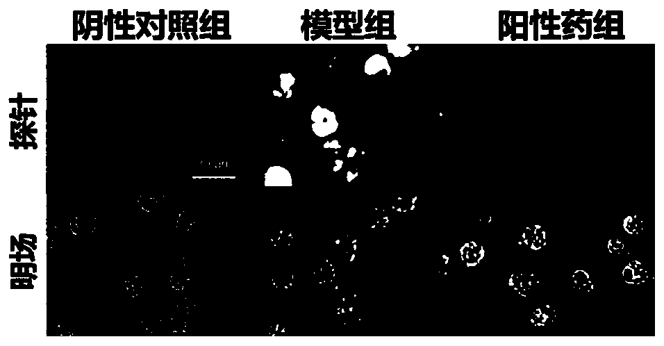
[0016] Example 1 Application of Formula I Molecules in Primary Alveolar Macrophage Staining

[0017] The 6-8 week C57BL / 6J mice were divided into three groups according to body weight: negative control group, model group and positive drug group. The model group and the positive drug cloth group were injected with 3 mg / kg bleomycin into the trachea to simulate human idiopathic pulmonary fibrosis, while the negative control group was given the same volume of normal saline as a negative control. The mice in the positive drug group were given the positive drug nintedanib 30mg / kg by oral gavage on the second day after the model was established for drug interference treatment, and the other two groups were given the corresponding solvent as a control. After 21 days of the entire model cycle, the mice were treated with Euthanasia.

[0018] The primary alveolar macrophages were isolated from the three groups of mice. After the culture was stable, the cells were transferred to a 96-we...

Embodiment 2

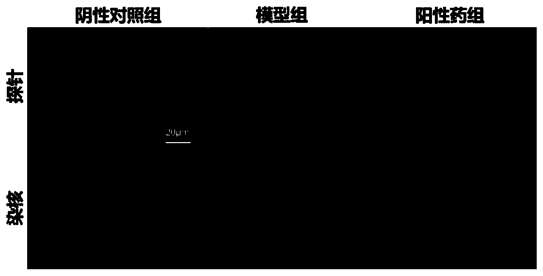
[0019] Application of embodiment 2 formula I molecule in lung tissue section staining

[0020] The mouse model is the same as described in Example 1. After the mouse is euthanized, the lungs of the mouse are taken out and fixed with 4% paraformaldehyde for 24 hours, and then the tissue is subjected to a series of dehydration, transparency, wax immersion, embedding, sectioning, and patching 1. Baking the slices to make paraffin sections, and then incubating the slices with 10 μM molecular probe of formula Ⅰ for 30 minutes, washing off with PBS, staining the nuclei with Hoechst reagent, washing off the PBS again, and collecting images with a fluorescence microscope. see results figure 2 , the tissue of the model group showed bright staining results, followed by the positive drug group, and the staining results of the negative control group were weaker, indicating that the probe shown in formula I can detect tissue damage caused by bleomycin modeling.

Embodiment 3

[0021] Example 3 Application of Molecules of Formula I in Live Mouse Imaging of Pulmonary Fibrosis.

[0022] The mouse model is the same as that described in Example 1. On the 21st day, after the mouse is shaved, the molecular probe of formula I with a concentration of 10 mg / kg is injected into the tail vein, anesthetized with isoflurane gas 1.5 hours later, and placed in a small Images were acquired with an In Vivo Imaging System (IVIS), and mice were euthanized at the end. see results image 3 , after bleomycin modeling, the mouse lung tissue showed a more obvious staining effect of probe I, indicating that the probe shown in formula I can detect pulmonary fibrosis caused by bleomycin modeling.


PUM


Abstract

The invention provides a fluorescence probe used for pulmonary fibrosis dyeing, and particularly relates to a micromolecule fluorescence probe for pulmonary fibrosis tissue or cell dyeing. The fluorescence probe provided by the invention has good stability, can be stored and used for a long time, is nontoxic and safe in use, has a high detection signal-to-noise ratio and high sensitivity, is simple and quick in operation, is high in result reproducibility, can be applied to cell, tissue and pathological process evaluation, and is especially applied to pulmonary fibrosis pathologic cell dyeing,pulmonary fibrosis tissue slice dyeing and pulmonary fibrosis model detection. The structure of the probe is disclosed in the following diagram.

Description

technical field [0001] The invention belongs to the field of biological detection, and relates to a fluorescent probe used for evaluating and staining pulmonary fibrosis pathological cells, tissues and pathological processes, in particular to a fluorescent probe used for pulmonary fibrosis staining and its application. [0002] technical background [0003] Pulmonary fibrosis is a type of interstitial disease that is caused by various causes of lung inflammation, persistent alveolar damage, and repeated destruction, repair, reconstruction, and excessive deposition of extracellular matrix, which eventually leads to changes in lung tissue structure and loss of function. The cause of most patients with pulmonary fibrosis is unknown, so it is called idiopathic pulmonary fibrosis (IPF). IPF is a "tumor-like disease" that mostly occurs in the elderly over 50 years old. The average survival period after diagnosis is only 2.8 years, and the mortality rate is higher than most tumors. ...

Claims


Application Information

Patent Timeline
Patent Type & Authority Applications(China)
IPC IPC(8): G01N21/64A61K49/00
CPCA61K49/0021G01N21/6428
Inventor 李新李佳胡永洲臧奕李晓荣董莹
Owner ZHEJIANG UNIV
Why Eureka
Try Eureka