ELISA detection method for canine distemper virus and antibody

A technology for canine distemper virus and canine distemper, applied in the direction of virus/bacteriophage, virus, virus peptide, etc., to achieve good antigenic effect

Inactive Publication Date: 2019-08-09
SOUTH CHINA AGRI UNIV
View PDF5 Cites 3 Cited by
  • Summary
  • Abstract
  • Description
  • Claims
  • Application Information

AI Technical Summary

Problems solved by technology

[0005] The purpose of the present invention is to overcome the deficiency that the method for detecting canine distemper antibody cannot be quantified in the existing market in the prior art, provide a kind of detection method of canine distemper antibody, as the effective detection method of quantitative detection canine distemper antibody content

Method used


Image

  • ELISA detection method for canine distemper virus and antibody
  • ELISA detection method for canine distemper virus and antibody
  • ELISA detection method for canine distemper virus and antibody

Examples


Embodiment 1

[0075] Example 1 Obtaining of pET-22b-N vector

[0076] 1. Experimental method

[0077] 1. Use vero cells to culture the CDV-1 virus strain, freeze and thaw it three times, and collect the virus. Use the small amount of RNA extraction kit to extract the virus RNA, and use the RNA inversion kit to reverse the extracted RNA into cDNA, and save it. to 4°C refrigerator.

[0078] 2. According to the CDVgp1nucleocapsid protein N gene sequence (Gene ID: 1489798) registered in GenBank, using the inverted cDNA (nucleotide sequence shown in SEQ IN NO: 1) as a template, design a pair of primers N-F / N-R ( Table 1), amplify the N coding region, introduce SalI and NotI restriction sites and His tag sequence, and obtain the N target fragment.

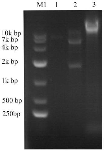
[0079] 3. Connect the N fragment and the pMD18-T vector by TA cloning, and then carry out transformation screening to obtain the recombinant plasmid pMD-N; the recombinant plasmids pMD-N and pET-22b are respectively treated with restriction enzymes ...

Embodiment 2

[0088] Example 2 Obtaining of CDV N protein

[0089] 1. Experimental method

[0090] 1. Transfer pET-22b-N from Example 1 or into E.coli DH5α competent, and after being identified as positive, extract the plasmid after massive expression in Amp-resistant LB.

[0091] 2. Transform the extracted pET-22b-N into Rosetta (DE3) competent bacteria, amplify the target bacterial strain, obtain the target plasmid pET-22b-N through the plasmid extraction kit, and transform it into the expression strain Rosetta (DE3 ) competence, plate screening to obtain a single clone of the target strain.

[0092] 3. Pick a single colony identified as positive and culture it to OD 600 When = 0.8 ~ 1, add IPTG to the medium to make the final concentration 1mmol / L, continue to cultivate for 6 hours, and collect the precipitate by centrifugation at 12000r / min for 15 minutes.

[0093] 4. Resuspend the bacterial cells induced in the previous step in PBS for sonication, and centrifuge at 12,000 r / min at 4...

Embodiment 3

[0114] Example 3 CDV N protein immunized mice

[0115] 1. Experimental method

[0116] 1. Use the BCA protein quantification method to determine the concentration of the expression product in Example 2, dilute the obtained protein with PBS to 100 μg / 0.1mL, and mix it with Freund’s complete adjuvant in equal volume; use the piston tube method to emulsify the target protein to make the protein Form a "water-in-oil" state. The judgment method is to drop the emulsified protein into a plate filled with water. If the target protein does not spread within 10 minutes, it means that the emulsification is successful; Inject 300 μg / mouse at multiple points on the back of the mouse, and inject 0.2 g / mL 200-400 mL of veterinary penicillin sodium into the leg muscles of the mice. The specific injection volume depends on the weight of the mice.

[0117] 2. The second immunization was carried out on the 14th day after the first step of immunizing the mice to strengthen the immune effect. Th...


PUM


Abstract

The invention discloses an ELISA detection method for a canine distemper virus and antibody. A canine distemper virus N protein is used to make a canine distemper detection kit, and the amino acid sequence is shown in SEQ ID NO:1. The canine distemper virus N protein is only used as an antibody to build the ELISA detection method, the N protein has good antigenicity as a canine distemper virus conserved protein and can quantify anti-canine distemper virus antibodies in a serum sample on the premise of preventing virus spread, the immune status of a blood-derived organism for the canine distemper virus or the immune level of the canine distemper virus secreted by a cell can be detected, more safety and higher efficiency are achieved compared with other products, and positive meaning is achieved for basic research and clinical detection on the canine distemper virus.

Description

technical field [0001] The invention relates to the technical fields of animal virology and animal immunity, and more specifically, relates to an ELISA detection method for canine distemper virus and antibodies. Background technique [0002] Canine distemper is a disease of canine animals. It belongs to the third category of animal diseases. It mainly infects canine animals. The transmission channel is mainly through direct contact with sick animals, and can also be transmitted through air or food. It is not a zoonotic disease. . [0003] Canine distemper is a highly contagious disease caused by canine distemper virus (CDV), which is highly contagious and has a mortality rate of up to more than 80%. In the early stage of canine distemper symptoms, the dog's body temperature was as high as 39.5 to 41 degrees Celsius, with loss of appetite, depression, watery secretions from eyes and noses, sneezing, and diarrhea. In the next 2 to 14 days, elevated body temperature, cough, p...

Claims


Application Information

Patent Timeline
IPC IPC(8): G01N33/68G01N33/569C07K14/12C12N15/45C12N15/70
CPCC07K14/005C12N15/70C12N2760/18422G01N33/56983G01N33/6854
Inventor 郭霄峰姜贺翟燕镅
Owner SOUTH CHINA AGRI UNIV
Why Eureka
Try Eureka