Binding protein and epitope-blocking ELISA for the universal detection of H5-subtype influenza viruses

a technology of h5-subtype influenza viruses and binding proteins, which is applied in the field of specific serological assays, can solve the problems of loss affecting the income of millions of people, limited use to clinicians, and loss of millions of dollars in poultry industry, and achieves stable and high-antigenic effects

Active Publication Date: 2013-11-05
TEMASEK LIFE SCIENCES LABORATORY
View PDF4 Cites 16 Cited by
  • Summary
  • Abstract
  • Description
  • Claims
  • Application Information

AI Technical Summary

Benefits of technology

[0012]In a further aspect, the invention comprises a method for detecting H5 subtype AIV in a biological specimen which comprises contacting the specimen with an antigen which contains an epitope of an H5 subtype hemagglutinin glycoprotein and determining whether an antibody in the specimen binds to the epitope. Preferably the binding determination is made in an epitope blocking ELISA. Preferably the epitope of the H5 subtype hemagglutinin glycoprotein includes the sequence CNTKCQTP (SEQ ID NO:1) or the sequence IHPLTIGE (SEQ ID NO:2). Both the IHPLTIGE (SEQ ID NO:2) and the CNTKCQTP (SEQ ID NO:1) epitopes exist in all known human H5N1 AIV strains, in essentially all known chicken H5N1 AIV strains, and in essentially all known H5N1 strains of other animals and birds, and are very stable and highly antigenic, making them very useful for diagnosing H5N1 infections.

Problems solved by technology

The poultry industry has lost millions of dollars to avian influenza.
This loss has affected the incomes of millions of people whose livelihoods depend on poultry.
Standard virus-isolation procedures have the disadvantage of requiring several days to obtain results, thereby making them of limited use to a clinician.
The disadvantages of RT-PCR include the high costs involved, the need for technically proficient staff, likelihood of contamination and the consequent risk of false positive results.
In addition, PCR primers may require constant updating because of antigenic drift [7].
However, neutralization assay and HI assay are not considered highly sensitive and necessitate further sub-typing and also are quite labor-intensive and time-consuming [8,9] and so are not ideal for large-scale routine testing of sera.
ELISA has been widely used as a pre-screening tool for investigating large numbers of samples, but indirect ELISA systems are commercially available only for chicken and turkey sera due to the unavailability of species-specific secondary antibodies of other species.
A further limitation of indirect ELISA is the need for high antigen purity.
The most significant disadvantage of the indirect ELISA, however, is that the HA antigen is known to cross-react with the other subtypes of viruses.
As a consequence, the indirect ELISA is not a dependable method for detection.
Most of these methods not only are cumbersome and labor intensive but also are time-consuming and include a risk of obtaining false positive results.
A further limitation of these techniques is that influenza viruses are segmented genome RNA viruses which are known to undergo continuous mutations and genetic re-assortments (antigenic drift), making it difficult to detect the virus [10].

Method used


Image

  • Binding protein and epitope-blocking ELISA for the universal detection of H5-subtype influenza viruses
  • Binding protein and epitope-blocking ELISA for the universal detection of H5-subtype influenza viruses
  • Binding protein and epitope-blocking ELISA for the universal detection of H5-subtype influenza viruses

Examples


example 1

I. Experimental

Viruses

[0053]Twenty four isolates of Indonesian H5N1 influenza strains used in this study and listed in Table 1 below (entries 1-24) were obtained from the National Institute of Health, Research and Development, Indonesia. Other H5 and non-H5 subtypes were provided by the Agri-Food and Veterinary Authority (AVA) of Singapore (entries 25-31 of Table 1).

[0054]

TABLE 1The avian influenza viruses used in this experimentSerial No.VirusesSubtypes1.Human / Indonesia / CDC7 / 06*H5N12.Human / Indonesia / CDC326 / 06*H5N13.Human / Indonesia / CDC329 / 06*H5N14.Human / Indonesia / CDC370 / 06*H5N15.Human / Indonesia / CDC390 / 06*H5N16.Human / Indonesia / CDC523 / 06*H5N17.Human / Indonesia / CDC594 / 06*H5N18.Human / Indonesia / CDC595 / 06*H5N19.Human / Indonesia / CDC597 / 06*H5N110.Human / Indonesia / CDC610 / 06*H5N111.Human / Indonesia / CDC623 / 06*H5N112.Human / Indonesia / CDC644 / 06*H5N113.Human / Indonesia / CDC669 / 06*H5N114.Human / Indonesia / TLL01 / 06*H5N115.Human / Indonesia / TLL02 / 06*H5N116.Duck / Indonesia / TLL60 / 06*H5N117.Human / Indonesia / TLL177 / ...

example 2

I. Experimental

[0083]A second monoclonal antibody, designated 1G5, was generated following the general procedures of Example 1 to obtain hybridoma supernatant. The mAb was raised against the recombinant HA0 protein, cloned from H5N1 influenza virus strain A / goose / Guangdong / 97.

Immunofluorescence Assay (IFA)

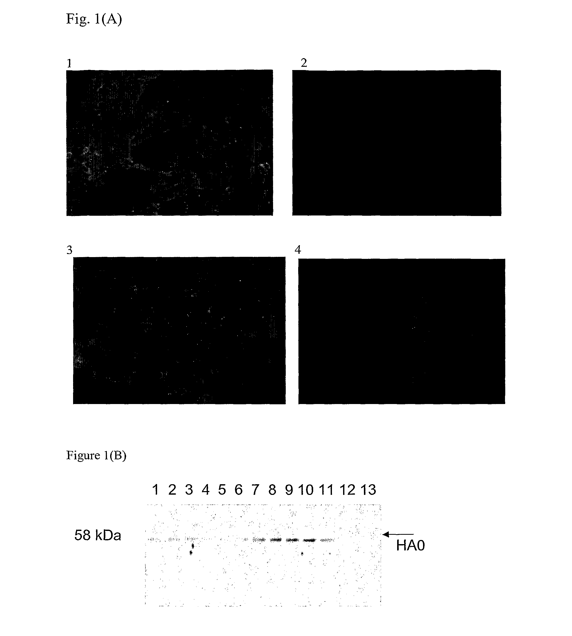
[0084]Sf-9 and MDCK cells were seeded in 960 well plates and incubated with recombinant baculovirus harboring truncated H5N1 HA1 gene and H5N1 viruses, respectively, At 36 hours post-infection, the cells were fixed with 4% paraformaldehyde for 30 minutes at room temperature. All washes were performed with PBS, pH 7.4. Fixed cells were incubated with hybridoma culture fluid at 37° C. for 1 hour, washed and incubated with a 1:50 dilution of fluorescein isothiocyanate (FITC)-conjugated rabbit anti-mouse Ig. Cells were washed and fluorescence was visualized using an Olympus IX71 microscope with appropriate barrier and excitation filters.

[0085]One mAb, designated 1G5, determined to be a...

example 3

Development of Antigen Capture ELISA (AC-ELISA)

[0101]96-well plates (Nunc, Denmark) were coated with purified mAbs 5F8 and / or 1G5 in 50 μl carbonate buffer (73 mM sodium bicarbonate and 30 mM sodium carbonate) and incubated at 37° C. for 1 hour or at 4° C. overnight. After each incubation step, the plates were washed 3 times with PBS containing 0.05% Tween 20 (PBST), and all dilutions were made in PBST containing 1% non-fat milk. The plates were blocked by incubation with 100 μl of blocking solution (5% non-fat milk in PBS-T at 37° C. for 1 hour, rinsed and incubated with 50 μl of purified highly pathogenic H5N1 Indonesian strains / low pathogenic AIV H5 subtypes (H5N1 / PR8, H5N2 and H5N3) / other non-H5 subtypes at 37° C. for 1 hour. After rinsing, 100 μl of guinea pig monospecific antibody IgG (1:500 dilution) were added, incubated for 1 hour at 37° C., washed and further incubated with 100 μl of HRP-conjugated rabbit anti-guinea pig immunoglobulin (diluted 1:1000). Color development w...


PUM

PropertyMeasurementUnit
volumeaaaaaaaaaa
pHaaaaaaaaaa
sizeaaaaaaaaaa

Abstract

Monoclonal antibodies and related binding proteins specific to influenza H5 subtype HA protein can be used in serological diagnosis of influenza H5 infection in mammalian and avian serum samples, including human serum samples. Each antibody reacts strongly with a wide variety of strains of H5 subtype and does not show cross-reactivity with non-H5 influenza subtypes.

Description

[0001]This application is a national filing under 35 USC §371 of PCT / SG2008 / 000043, filed Feb. 5, 2008. The prior application is incorporated herein.SEQUENCE SUBMISSION[0002]The present application includes a Sequence Listing filed in electronic format. The Sequence Listing is entitled 2577185SequenceListing.txt, was created on 8 Jan. 2013 and is 2 kb in size. The information in the electronic format of the Sequence Listing is part of the present application and is incorporated herein by reference in its entirety.FIELD OF THE INVENTION[0003]This invention relates to the development of a specific serological assay for the detection of antibodies to avian influenza viruses. More specifically, this invention relates to the production of monoclonal antibodies which react strongly with all known subtypes of H5 influenza virus, but which do not cross-react with non-H5 influenza subtypes, and are used to develop epitope blocking ELISAs to detect serum antibody and related binding protein t...

Claims


Application Information

Patent Timeline
Patent Type & Authority Patents(United States)
IPC IPC(8): C12Q1/70C07K16/10
CPCC07K16/1018G01N33/56983G01N2333/11C07K2317/34
Inventor MOOKKAN, PRABAKARANPADUBIDHRI, NAYANA PRABHUVELUMANI, SUMATHYKWANG, HWEI-SING JIMMY
Owner TEMASEK LIFE SCIENCES LABORATORY
Why Eureka
Try Eureka