SERS-immunochromatography detection method for rapidly and highly sensitively detecting Mycoplasma pneumoniae infection

A technology for immunochromatographic detection and mycoplasma pneumoniae, which is applied in measuring devices, analytical materials, biological testing, etc., can solve the problems of low detection sensitivity, achieve high sensitivity, simple detection process, and improve detection sensitivity

Inactive Publication Date: 2019-08-16
ACADEMY OF MILITARY MEDICAL SCI
View PDF14 Cites 8 Cited by
  • Summary
  • Abstract
  • Description
  • Claims
  • Application Information

AI Technical Summary

Problems solved by technology

[0004] The present invention is aimed at the low problem of detection sensitivity of traditional colloidal gold immunochromatography technology, discloses the preparation method of SERS-...

Method used


Image

  • SERS-immunochromatography detection method for rapidly and highly sensitively detecting Mycoplasma pneumoniae infection
  • SERS-immunochromatography detection method for rapidly and highly sensitively detecting Mycoplasma pneumoniae infection
  • SERS-immunochromatography detection method for rapidly and highly sensitively detecting Mycoplasma pneumoniae infection

Examples


Embodiment 1

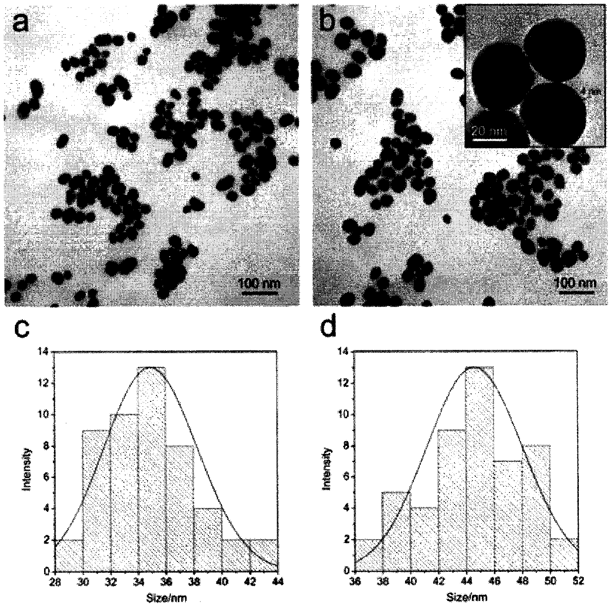
[0038] Example 1, Characterization of Au / DTNB@Ag / DTNB SERS probe

[0039] Experimental procedure: In this experiment, 1.5mL of 1wt% sodium citrate solution was added to 100mL of boiling 0.01wt% chloroauric acid solution, kept boiling for 15 minutes, and then cooled to room temperature. The particle size of AuNPs prepared by this parameter is about 35nm. 10 μL of 10 mM DTNB ethanol solution was added to 10 mL of AuNPs solution, and the mixture was stirred at room temperature for 4 hours. Centrifuge at 8000 rpm for 8 minutes to remove excess DTNB molecules, resuspend the pellet in 10 mL of deionized water, dilute 10 mL of Au NPs / DTNB solution fourfold with deionized water and boil. Subsequently, 0.5 mL of sodium citrate (1%, w / v) was added to the solution and stirred. Finally, 1 mL of silver nitrate (1 mM) was added dropwise to the above solution, stirred and boiled for 15 minutes. At this stage, the solution of Au NPs turned orange-yellow, indicating the formation of Au / DTNB...

Embodiment 2

[0041] Embodiment 2, SERS-immunochromatography test strip quantitatively detects and analyzes the result of human IgM

[0042] Experimental procedure: Dilute human IgM sequentially to 1000ng mL -1 , 100ng mL -1 , 10ng mL -1 , 1ng mL -1 , 0.5ng mL -1 and 0.1ng mL -1 A total of six concentrations, 1×pBST (1×PBS, 0.05% Tween-20) as a blank control; take 70 μL of the sample solution and add it to the sample area of ​​the SERS-immunochromatography test strip in turn, and use Raman after 15 minutes The spectrometer detects the Raman signal in the detection zone. Draw Raman Peak 1335cm -1 SERS signal intensity and human IgM concentration (0.1-1000ngmL -1 ) calibration curve between logarithms. at 1335cm -1 Draw the SERS spectrum in an extended area (250 μm × 50 μm) with a step size of 5 μm to obtain the uniformity result of the SERS signal on the detection line.

[0043] Experimental results: When the concentration of human IgM increases, due to the formation of many immune...

Embodiment 3

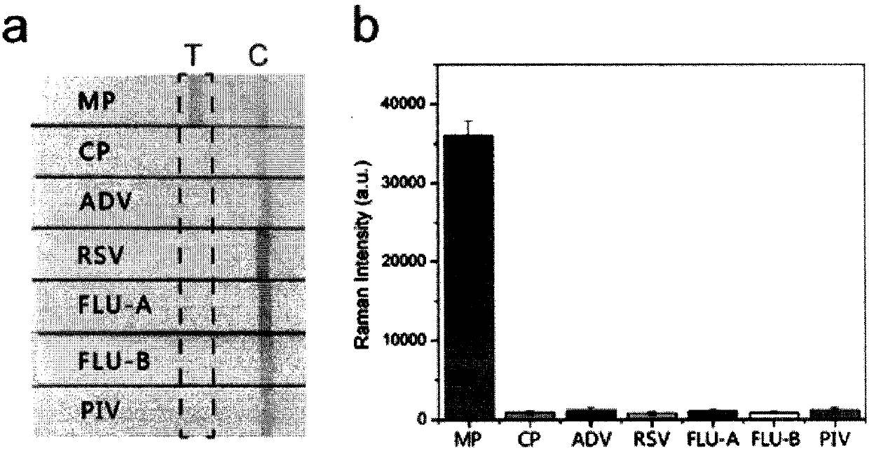
[0044] Embodiment 3, SERS immunochromatographic test strip detects the result of 20 routine pulmonary branch IgM positive serum specimens

[0045] Experimental procedure: 20 cases of pulmonary branch-specific IgM-positive clinical serum samples and 10 cases of pulmonary branch-specific IgM-negative clinical serum samples were detected with SERS-immunochromatographic test strips. After mixing 10 μL of clinical serum and 60 μL of sodium chloride solution (w / v), it was added dropwise to the sample pad of the prepared SERS-immunochromatography test strip, and the result was detected after 15 minutes. Based on the immune reaction and chromatography, the lung branch-specific IgM-positive serum samples will form two red detection lines and control lines visible to the naked eye, and the lung branch-specific IgM-negative serum samples will only form a red control line visible to the naked eye . Detection and analysis come from SERS-immunochromatography test strip detection line 1335c...


PUM

PropertyMeasurementUnit
Particle sizeaaaaaaaaaa
The average particle sizeaaaaaaaaaa

Abstract

The invention relates to a SERS-immunochromatography detection method for rapidly and highly sensitively detecting Mycoplasma pneumoniae infection. The detection principle is that Nitrocellulose membranes (NC) are used as a carrier, a double-layer dye 5,5'-dithiobis (2-nitrobenzoic acid){5,5'-dithiobis- (2-nitrozoic acid), DTNB} labeled Au@Ag nano material coupling detection antibody is used as anSERS probe, a novel surface enhanced Raman spectroscopy (SERS-ICA)-based immunochromatography technology is established by combining the traditional Immunochromatography assay (ICA), and the technology is used for detecting human IgM and Mycoplasma pneumoniae (MP) specific IgM positive serum samples. The detection comprises the processes of sample dilution, SERS probe release, antigen-antibody reaction, result analysis and the like. The SERS-immunochromatography detection method for rapidly and highly sensitively detecting Mycoplasma pneumoniae infection can improve the detection sensitivityof human IgM and the detection rate of the lung branch positive serum specimen, and has important significance for clinical treatment.

Description

technical field [0001] The invention relates to the technical field of in vitro diagnostic reagents, in particular to a SERS-immunochromatographic detection method for rapid and highly sensitive detection of Mycoplasma pneumoniae infection. Background technique [0002] Mycoplasma pneumoniae is a pathogen that lacks peptidoglycan cell walls, does not require host cells, and can replicate autonomously; it is prevalent in all age groups worldwide and is a major cause of pneumonia and respiratory diseases. Nearly 6%-30% of adults and 20%-40% of children suffer from community-acquired pneumonia due to infection with Mycoplasma pneumoniae, and there is an epidemic peak every 3-7 years. Antibody therapy is crucial. The existing gold standard method remains the traditional cell culture as the most reliable and cheap diagnostic method of lung bronchus. However, branch culture is time-consuming, requiring 2-8 weeks, and requires special dishes and specialized technicians. Another ...

Claims


Application Information

Patent Timeline
IPC IPC(8): G01N33/558G01N33/68
CPCG01N33/558G01N33/6854
Inventor 王升启贾小飞肖瑞汪崇文荣振
Owner ACADEMY OF MILITARY MEDICAL SCI
Why Eureka
Try Eureka