Blood biomarker for detecting uremia and application thereof

A biomarker, uremia technology, applied in the field of medicine, to achieve the effect of easy-to-learn operation steps, simple operation steps, and less dosage

Inactive Publication Date: 2019-10-18
THE AFFILIATED HOSPITAL OF QINGDAO UNIV
View PDF0 Cites 0 Cited by
  • Summary
  • Abstract
  • Description
  • Claims
  • Application Information

AI Technical Summary

Problems solved by technology

However, the CEM Discover single-mode microwave protein hydrolysis instrument introduced in this patent combines the advantages of high-efficiency microwave energy and the vacuum/gas exchange po

Method used


Image

  • Blood biomarker for detecting uremia and application thereof
  • Blood biomarker for detecting uremia and application thereof
  • Blood biomarker for detecting uremia and application thereof

Examples


Embodiment 1

[0049] 1. Effects of different acids on serum microwave acid hydrolysis

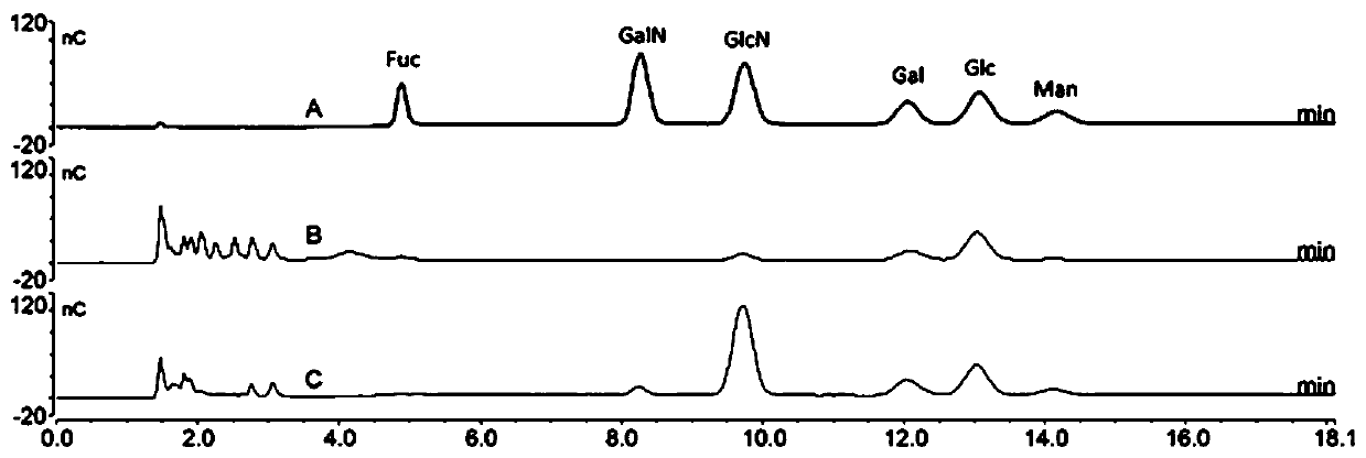
[0050] (1) Randomly take serum from 10 patients, take 2 parts for each serum, take 2 μL respectively to microwave hydrolysis tube, add deionized water to 10 μL, mix well, then add 10 μL 6mol / L HCl or 4mol / L TFA;

[0051] (2) Under the conditions of power 100w and temperature 100°C, use a single-mode microwave protein hydrolysis instrument to carry out microwave acid hydrolysis for 10 minutes;

[0052] (3) The sample after microwave acid hydrolysis was transferred to a 1.5mL centrifuge tube with 20 μL of deionized water, repeated three times, and concentrated by centrifugation to remove acid;

[0053] (4) Add 100 μL of methanol to each tube, centrifuge and concentrate to remove residual HCl, repeat three times;

[0054](5) Each tube of the obtained dried sample was dissolved in 150 μL of deionized water at a speed of 13000 r / min, centrifuged for 5 min, and the supernatant was taken for anion exchange chr...

Embodiment 2

[0075] 1. Drawing of monosaccharide standard curve

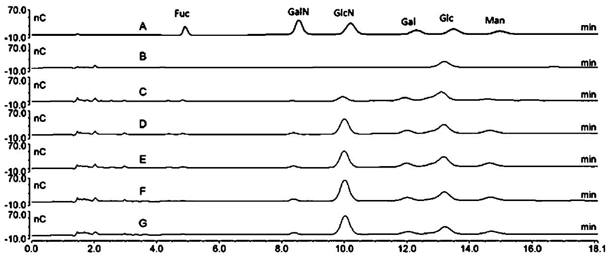
[0076] Accurately weigh the appropriate amount of fucose, galactosamine, glucosamine, galactose, glucose and mannose, add deionized water to prepare the above monosaccharides containing 0.05mg / mL, 0.01mg / mL, 0.005mg / mL, 0.001 mg / mL, 0.0005mg / mL, 0.0001mg / mL, 0.00005mg / mL solution; take 80μL to the sample bottle for anion exchange chromatography analysis.

[0077] The chromatographic conditions are as follows:

[0078] Analytical column: Thermo Scientific Dionex Carbo PAC TM PA10, 4.0mm×250mm;

[0079] Guard column: Thermo Scientific Dionex Carbo PAC TM PA10, 4.0mm×50mm;

[0080] Eluent: use 18mM NaOH for 0-18min; flow rate: 1.0mL / min, injection volume: 10μL, column temperature: 30℃,

[0081] Detector: electrochemical detector, gold electrode (P / N 061875), standard sugar potential, running time: 18min.

[0082] Anion-exchange chromatograms of six monosaccharide standards were obtained, see Figure 6 a. From Figure...

Embodiment 3

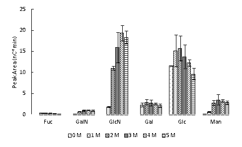
[0095] Get 2 μ L of blood from 216 confirmed uremia patients. The microwave acid hydrolysis and ion chromatography detection steps of the blood are the same as those in Example 1. For the statistical detection results, see Figure 7 .

[0096] Will Figure 7 The results are summarized in Table 1, and the analysis results of the six monosaccharide concentrations are shown in Table 2.

[0097] Table 1. The relative trend of the 6 monosaccharides obtained after blood degradation in patients with uremia and those without uremia

[0098] Fucose Galactosamine Glucosamine Galactose glucose Mannose Uremia ↑* ↑*** ↑*** NS ↑* NS

[0099] Note: "NS" in the population without uremia means no change compared with the population without uremia, "***p<0.001", "**p<0.01" and "*p<0.05" represent the Compared with the uremic population, there are different significant differences, "↑" and "↓" respectively represent the increase or decrease compared with...


PUM


Abstract

The invention provides a blood biomarker for detecting uremia and application thereof. The biomarker includes galactosamine and glucosamine that are obtained by processing blood based on microwave acid hydrolysis and anion exchange chromatography with pulsed amperometric detection. The degradation is fast, the batch throughput and compatibility are high, the acid consumption is reduced, and the operation steps are simple and are easy to learn; and the sensitivity and accuracy are high. The obtained results show that the degradation analysis method can rapidly degrade the blood sample, analyzethe monosaccharide composition in the blood sample, and calculate the concentration of the monosaccharide; and the person without uremia and a patient with uremia can be distinguished to some extents.

Description

technical field [0001] The invention belongs to the field of medicine, and in particular relates to a biomarker for detecting uremia and its application. Background technique [0002] The kidney is an important organ of the urinary system. Its main function is to produce and excrete urine, and to excrete the metabolic waste of the human body. generation and bone growth etc. Therefore, kidney disease has a great impact on the function of the human body. End-stage renal disease often develops into uremia. Renal replacement therapy (peritoneal dialysis, hemodialysis, kidney transplantation) is the only effective treatment for end-stage renal disease, but dialysis has a higher risk of infection from other diseases. Therefore, it is particularly important to find an early diagnosis method. [0003] Diagnostic methods for kidney disease include urinalysis (proteinuria, hematuria, casts, and leukocyturia), measurement of glomerular filtration rate, imaging studies, and renal bi...

Claims


Application Information

Patent Timeline
IPC IPC(8): G01N30/96G01N30/06
CPCG01N30/96G01N30/06
Inventor 张丽娟何燕利
Owner THE AFFILIATED HOSPITAL OF QINGDAO UNIV
Why Eureka
Try Eureka