
Method for preparing tissue engineering tendon materials by ultrasonic concussion technology

An ultrasonic vibration and tendon technology, applied in tissue regeneration, medical science, prosthesis, etc., can solve the problems of limiting the application of acellular tendons, unfavorable cell adhesion, migration and proliferation, etc., and achieves small damage, good biomechanical properties, low impact effect

Pending Publication Date: 2019-10-29
SHANGHAI CHANGZHENG HOSPITAL
View PDF5 Cites 4 Cited by
  • Summary
  • Abstract
  • Description
  • Claims
  • Application Information

AI Technical Summary

Problems solved by technology

Therefore, the acellular matrix obtained by the existing decellularization method is not conducive to cell adhesion, migration and proliferation, these factors limit the application of decellularized tendon in the field of tissue engineering tendon

Method used


Image

  • Method for preparing tissue engineering tendon materials by ultrasonic concussion technology
  • Method for preparing tissue engineering tendon materials by ultrasonic concussion technology
  • Method for preparing tissue engineering tendon materials by ultrasonic concussion technology

Examples


Embodiment 1

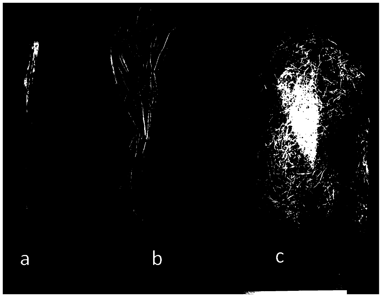
[0033] Example 1: Ultrasonic vibration technology is used in the preparation of bovine tendon acellular matrix:

[0034] 1) Take fresh bovine tendon / ligament, and remove the adventitia and synovial tissue on the surface of the tendon / ligament.

[0035] 2) Place the tendon / ligament in a vacuum freeze dryer at -56°C for 48 hours.

[0036] 3) The tendon / ligament structure is loosened by drying at low temperature through physical loosening methods such as extrusion, hammering, vacuum anti-explosion airflow impact and other methods.

[0037] 4) The loose tendon / ligament is further physically and mechanically combed to separate the tendon / ligament collagen fibers or collagen fiber bundles.

[0038] 5) Put the isolated collagen fibers or fiber bundles into 10mM hypotonic Tris solution, add 0.05% serine protease inhibitor (0.05% phenylmethylsulfonyl fluoride ethanol solvent, 35ml / L 50ml ethanol + 25mgPMSF), at room temperature The shaker was shaken for 12h. Pour off the solution, a...

Embodiment 2

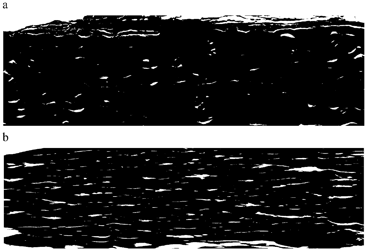
[0041] Example 2: Histological examination

[0042] Collagen fibers or fiber bundles before and after decellularization were taken for paraffin embedding, sectioned (5 μm), and HE stained for observation. Observe the cell nucleus components (hematoxylin staining), collagen arrangement (eosin staining), and see that the cell components are completely removed ( figure 2 ).

Embodiment 3

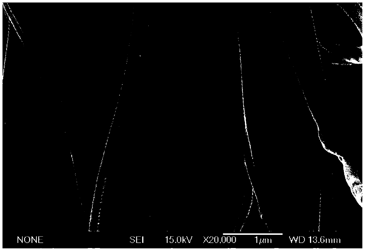
[0043] Embodiment 3: Electron microscope inspection

[0044] Cut the tendon acellular matrix into a length of about 5mm for later use, take the loading plate, fix the freeze-dried fresh tendon and freeze-dried tendon fiber bundle samples on the surface of the loading plate, spray gold powder, put it into the scanning electron microscope, and extract the internal air to be in a vacuum state , start detection. The results showed that the decellularization of tendon into three-dimensional helical structure did not change ( image 3 ).


PUM


Abstract

The invention relates to the technical field of tissue engineering decellularization, in particular to a method for preparing tendon / ligament acellular matrix materials by an ultrasonic concussion technology. According to the method, the ultrasonic concussion technology is used for decellularization, mechanical structure units and cell attachment structures inherent in the tendon collagen fibers or fiber bundles are retained, and the good mechanical performance and the good biocompatibility are achieved. A tendon extracellular matrix decellularized by the ultrasonic concussion technology is used, in addition to application of repair and reconstruction of the ligament and the tendon, the tendon extracellular matrix can be further widely applied to other biomedical fields since the characteristics of a fibrous material is achieved, and the social required quantity is great.

Description

technical field [0001] The invention relates to the technical field of preparation of tissue engineering biological materials, in particular to a new method for decellularizing tissue engineering tendon materials by using ultrasonic vibration technology. Background technique [0002] Tendons and ligaments have poor self-healing ability after injury, leading to fibrous scar formation, affecting limb activities, reducing the stress of tendons and ligaments, and increasing the risk of re-injury. Therefore, the repair and reconstruction of tendon and ligament defects are very important to improve the quality of life of patients. At present, the main methods for repairing tendons and ligaments include autograft and allograft. Autograft is currently the most effective treatment for tendon and ligament reconstruction. However, it can cause damage to the donor site, and the source is limited; there is immune rejection in allogeneic transplantation; synthetic materials such as carbo...

Claims


Application Information

Patent Timeline
IPC IPC(8): A61L27/36A61L27/50
CPCA61L27/3633A61L27/3662A61L27/3691A61L27/50A61L2430/10A61L2430/40
Inventor 陈雄生周盛源黄文茂苑博唐一钒王智巍
Owner SHANGHAI CHANGZHENG HOSPITAL
Learn More