Bioactivity bracket and preparation method thereof

A bioactive, composite stent technology, applied in the field of bioactive stents and their preparation, achieves the effects of high pore volume and specific surface area, improving drug activity, and improving drug encapsulation efficiency

Active Publication Date: 2019-11-12
SICHUAN UNIV
View PDF8 Cites 14 Cited by
  • Summary
  • Abstract
  • Description
  • Claims
  • Application Information

AI Technical Summary

Problems solved by technology

The use of hollow mesoporous hydroxyapatite surface amination treatment to lo

Method used


Image

  • Bioactivity bracket and preparation method thereof
  • Bioactivity bracket and preparation method thereof
  • Bioactivity bracket and preparation method thereof

Examples


Embodiment 1

[0043] A method for preparing a bioactive scaffold, comprising the following steps:

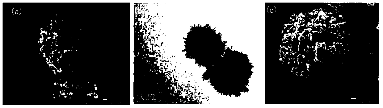
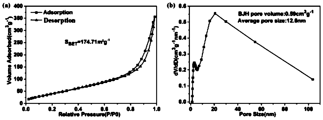
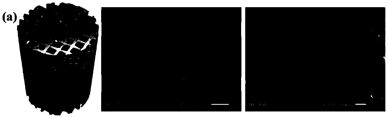
[0044] S1: Preparation of hollow mesoporous hydroxyapatite microspheres (MHA)

[0045] Add 1.963g creatine phosphate to 100mL distilled water to prepare A solution, add 1.225g calcium chloride dihydrate to 250mL distilled water to prepare B solution; take 10mL A solution and slowly drop into 30mL B solution, use 1mol / L Adjust the pH to about 10 with NaOH, and stir it with magnetic force for 30 minutes after the addition is complete; then move it to a microwave reactor, control the reaction temperature at 120°C, and microwave power at 5W, and continue to react for 30 minutes; then centrifuge, wash, and freeze-dry Get MHA.

[0046] S2: MHA surface functional modification and load bone morphogenetic protein

[0047] SS1: Soak MHA in absolute ethanol and ultrasonically disperse for 30min, then mix the dispersion with 20mmol / L γ-aminopropyltriethoxysilane (APS) ethanol solution at a volume ratio...

Embodiment 2

[0053] A method for preparing a bioactive scaffold, comprising the following steps:

[0054] S1: Preparation of hollow mesoporous hydroxyapatite microspheres (MHA)

[0055] Add 1.812g creatine phosphate to 100mL distilled water to prepare A solution, add 1.025g calcium chloride dihydrate to 250mL distilled water to prepare B solution; take 10mL A solution and slowly drop into 30mL B solution, use 1mol / L Adjust the pH to about 10 with NaOH, and stir it with magnetic force for 30 minutes after the addition is complete; then move it to a microwave reactor, control the reaction temperature to 150°C, and microwave power to 5W, and continue to react for 10 minutes; then centrifuge, wash, and freeze-dry Get MHA.

[0056] S2: MHA-loaded bone morphogenetic protein

[0057] Take 5 mg of the above-mentioned synthesized MHA, immerse in 50 mL of BMP-2 solution with a concentration of 0.2 μg / mL under aseptic conditions and oscillate at room temperature for equilibrium adsorption for 24 ho...

Embodiment 3

[0061] A method for preparing a bioactive scaffold, comprising the following steps:

[0062] S1: Preparation of hollow mesoporous hydroxyapatite microspheres (MHA)

[0063] Add 1.963g creatine phosphate to 100mL distilled water to prepare A solution, add 1.225g calcium chloride dihydrate to 250mL distilled water to prepare B solution; take 10mL A solution and slowly drop into 30mL B solution, use 1mol / L Adjust the pH to about 10 with NaOH, and stir the reaction for 30 minutes after the addition is complete; then move it to a microwave reactor, control the reaction temperature to 110°C, and the microwave power to 5W, and continue to react for 10 minutes; then centrifuge, wash, and freeze-dry Get MHA.

[0064] S2: MHA-loaded bone morphogenetic protein

[0065] Take 5 mg of the above-mentioned synthesized MHA, immerse in 50 mL of BMP-4 solution with a concentration of 0.3 μg / mL under aseptic conditions and oscillate at room temperature for equilibrium adsorption for 24 hours; ...


PUM

PropertyMeasurementUnit
Apertureaaaaaaaaaa
Apertureaaaaaaaaaa
Concentrationaaaaaaaaaa

Abstract

The invention discloses a bioactivity bracket and a preparation method thereof. The bioactivity bracket is obtained through the following steps of firstly, loading bone shape generation proteins on ahollow mesoporous hydroxylapatite microsphere through a grafting manner or an adsorbing manner, so as to obtain a drug bearing microsphere, then mixing the drug bearing microsphere into a bracket matrix solution, then performing printing through a 3D printing technique to prepare a compound bracket, and finally, adsorbing chemotactic growth factors on the surface to obtain the bioactivity bracket;or firstly, blending the drug bearing microsphere and the chemotactic growth factors in the bracket matrix solution, and then performing printing through the 3D printing technique to prepare the bioactivity bracket. Through the adoption of the method disclosed by the invention, the obtained bioactivity bracket can realize time sequence releasing of various growth factors: the chemotactic growth factors are released in an early stage, and bone repair cells are recruited to a damaging region; and bone shape generation proteins are mainly released in the later stage, and bone marrow substrate stem cells which are recruited to the damaging region are induced to be subjected to osteogenic differentiation, so that regeneration and repairing potential of an autologous tissue is sufficiently mobilized, and regeneration and repairing of bone defect are promoted.

Description

technical field [0001] The invention belongs to the technical field of biological scaffold preparation, and in particular relates to a bioactive scaffold and a preparation method thereof. Background technique [0002] Scaffold material is the most basic material and structural basis in bone tissue engineering. It can play an important role in the adhesion, proliferation, differentiation, material exchange and other processes of seed cells after implantation in the body, greatly affecting cell behavior and organization. reconstruction. In recent years, 3D printing technology has become a hotspot in the field and application of biomedicine. 3D printing technology manufactures scaffolds through layer-by-layer printing, which can accurately simulate the complex three-dimensional microstructure and scaffold shape of natural tissues, accurately control the distribution and spatial direction of micropores, and prepare scaffolds with controllable structure and highly interpenetrati...

Claims


Application Information

Patent Timeline
IPC IPC(8): A61L27/12A61L27/22A61L27/18A61L27/20A61L27/50A61L27/54A61L27/56A61L27/58B33Y10/00B33Y70/00
CPCA61L27/12A61L27/18A61L27/20A61L27/227A61L27/50A61L27/54A61L27/56A61L27/58A61L2300/252A61L2300/412A61L2300/414A61L2300/602A61L2430/02B33Y10/00B33Y70/00C08L89/00C08L67/04C08L5/08
Inventor 李吉东魏加伟李玉宝左奕邹琴
Owner SICHUAN UNIV
Why Eureka
Try Eureka