Construction method of liver unit stent, liver unit stent and application of liver unit stent in field of drug testing

A construction method and biological technology, applied in drug screening, artificial cell constructs, biochemical equipment and methods, etc., can solve problems such as inability to flexibly change, fixed liver tissue structure, and inability to reach engineering levels

Inactive Publication Date: 2019-11-29
REGENOVO BIOTECH
View PDF6 Cites 3 Cited by
  • Summary
  • Abstract
  • Description
  • Claims
  • Application Information

AI Technical Summary

Problems solved by technology

However, this method is limited to cells and cannot reach the engineering level; the constructed liver tissue s

Method used


Image

  • Construction method of liver unit stent, liver unit stent and application of liver unit stent in field of drug testing
  • Construction method of liver unit stent, liver unit stent and application of liver unit stent in field of drug testing
  • Construction method of liver unit stent, liver unit stent and application of liver unit stent in field of drug testing

Examples


Embodiment 1

[0065] Example 1: Preparation of Liver Unit Scaffold

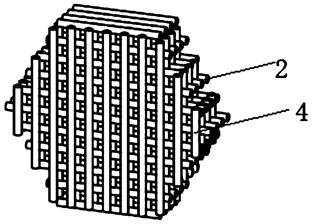
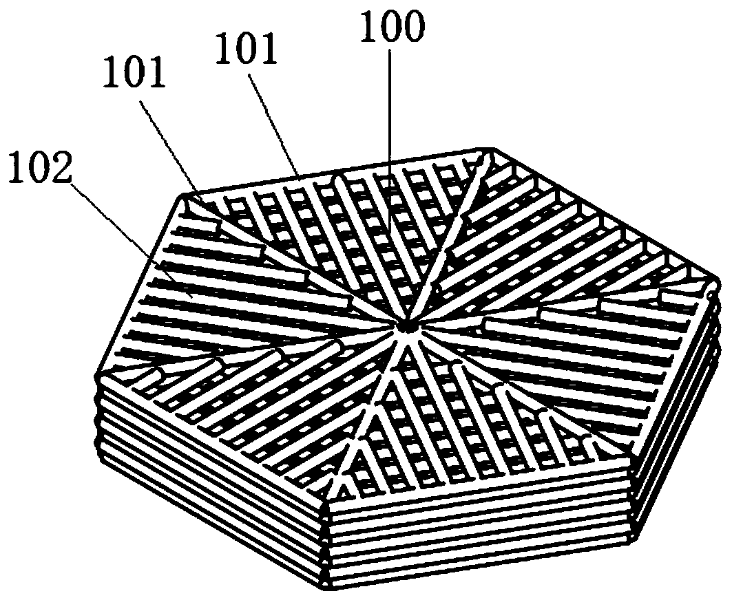
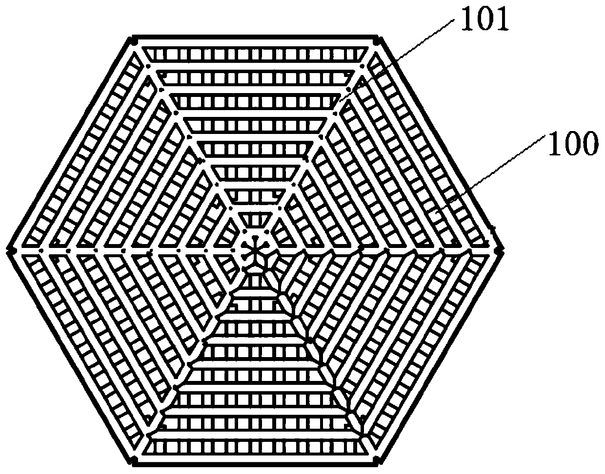
[0066] The preparation method of the liver unit scaffold comprises the following steps: (1) mixing gelatin monomer and solvent water at a mass ratio of 10:100 to prepare a gelatin solution, and mixing sodium alginate monomer and solvent water at a mass ratio of 2:100 to prepare Sodium alginate solution; (2) Digest and centrifuge the C3A cells cultured to the fusion stage, resuspend the cells in the medium, and prepare a single cell suspension; Mix the volume ratio to form a composition; (3) inhale the mixed hydrogel cell composition into a microsyringe, place it at 4°C for 1 hour, and assemble it on a 3D printer; (4) design the internal channel of the liver unit, the internal When filling, the nozzles are filled and printed at equal intervals along the first direction, and then the printing platform is lowered by 0.1mm along the Z axis, and the nozzles are filled and printed at equal intervals along the second direction pe...

Embodiment 2

[0070] Example 2: Construction of liver units containing cholangiocytes

[0071] The method of this embodiment includes the following steps: (1) mixing collagen monomer and solvent water at a mass ratio of 1:100 to prepare a collagen solution, and mixing sodium alginate monomer and solvent water at a mass ratio of 2:100 to prepare a collagen solution Sodium alginate solution; (2) Digest and centrifuge the HepaRG cells cultured to the fusion stage, resuspend the cells in the culture medium, and prepare a single cell suspension; press the cell suspension, collagen solution, sodium alginate and cytokine solution at a ratio of 0.05:5 : Mix at a volume ratio of 5:0.05 to form a composition, wherein the cytokine is a mixture of hepatocyte growth factor and insulin-like growth factor, wherein the volume ratio of hepatocyte growth factor and insulin-like growth factor in the mixture is 1 : 1; (3) Inhale the mixed hydrogel cell composition into a micro-syringe, place it at 4°C for 1 ho...

Embodiment 3

[0092] Example 3, the construction method of the liver unit scaffold kit is: (1) gelatin monomer and solvent water are mixed in a mass ratio of 10:100 to prepare a gelatin solution, and sodium alginate monomer and solvent water are mixed in a mass ratio of 2: 100 mixed to prepare a sodium alginate solution; (2) digest and centrifuge the C3A cells cultured to the fusion stage, resuspend the cells in the medium, and prepare a single cell suspension; press the cell suspension, gelatin solution and sodium alginate solution at 1: Mix at a mass ratio of 5:5 to form a composition; (3) Inhale the mixed hydrogel cell composition into a microinjector, place it at 4°C for 1 hour, and assemble it on a 3D printer; (4) Inside the liver unit Channel design, the first layer of nozzles prints the bracket along the first direction. After the first layer, the nozzle fills the bracket along the second direction perpendicular to the first direction, that is, the printing direction is rotated 90°, a...


PUM

PropertyMeasurementUnit
Diameteraaaaaaaaaa
Diameteraaaaaaaaaa
Viscosityaaaaaaaaaa

Abstract

The invention relates to the field of biomedicine, in particular to a construction method of a liver unit stent, a liver unit stent and application of the liver unit stent in the field of drug testing. The invention aims to provide a construction method of a liver unit stent, a liver unit stent and application of the liver unit stent in the field of drug testing. The construction method of the liver unit stent comprises the following steps: a gelled first composition, which includes hepatic parenchymatous cells and a biodegradable material, is loaded into a biological 3D printer, and an intermediate is printed according to a preset path; a cross-linking agent is dropwise added to the intermediate such that the biodegradable material in the intermediate is cross-linked to form a liver unitstent, wherein a channel is formed in the liver unit stent. In the liver unit stent, cell variety, quantity and position are controllable and the channel is controllable, thus providing a microenvironment for different cells. In addition, the liver unit stent can be used for researching the drug hepatotoxicity of new drugs and is used for detecting drug metabolites, gene expression, protein expression, cell apoptosis and cell proliferation.

Description

technical field [0001] The invention relates to the field of biomedicine, in particular to a method for constructing a liver unit scaffold, a liver unit scaffold and its application in the field of drug detection. Background technique [0002] Drug-induced liver disease refers to liver function damage caused by liver exposure to drugs without other infectious factors, and is one of the common causes of liver biochemical abnormalities. Drug-induced liver disease has become an increasingly serious problem in society due to the drugs that people take, and various nutritional and health products with drug functions, special foods, alcohol, etc. that people come into contact with in their daily life. Therefore, in order to reduce drug-induced liver disease, pharmaceutical companies often invest huge R&D expenses to screen candidate drugs. The average R&D expense for each approved new drug is about US$500-2 billion. [0003] The engineered liver tissue, its array and its preparat...

Claims


Application Information

Patent Timeline
IPC IPC(8): C12N5/071C12Q1/02
CPCC12N5/067C12N2503/02C12N2513/00G01N33/5067G01N2500/10
Inventor 徐铭恩周永勇王玲欧阳杨
Owner REGENOVO BIOTECH
Why Eureka
Try Eureka