

Preparation method of human skin stem cell factor nano-liposome-exosome complex
A nano-liposome and skin stem cell technology, applied in the field of cell biology, can solve problems such as difficult to reach target tissue, cause immune response, weak binding force of target tissue cells, etc., and achieve good stability
- Summary
- Abstract
- Description
- Claims
- Application Information
AI Technical Summary
Problems solved by technology
Method used
Image

Examples
Embodiment 1
[0049] The preparation of the nanoliposome of embodiment 1 coating cytokines
[0050] 1. Materials and steps
[0051] 1. Isolation of skin cells
[0052] Disinfection treatment of human skin tissue: Use a scalpel or scissors to remove the subcutaneous tissue (remove as clean as possible), wash once in 70% ethanol, and transfer the tissue to a PBS culture dish containing 2× double antibody for 3 min / time* 2 times;
[0053] The tissue was cut into 2mm*2mm, and digested with an appropriate amount of 0.1-5mg / ml Dispase in a 4°C refrigerator overnight; the next morning, the epidermis and dermis were separated with tweezers.
[0054] 2. Obtain primary skin epidermal stem cells
[0055] Add 10ml of 0.05% trypsin to the collected epidermis and digest it in a water bath at 37°C until a large number of cells fall off. Repeatedly pipette with DMEM medium containing 10% FBS for more than 20 times to obtain a single cell suspension, filter with a 100 micron filter, 1000rpm Centrifuge f...
Embodiment 2
[0075] Example 2 Obtaining of exosomes
[0076] 1. Materials and methods
[0077] (1) Preparation of exosome-free cell culture medium:
[0078] Centrifuge at 100,000g in an ultra-high-speed centrifuge at 4°C for 8 hours, collect the supernatant, and remove the exosomes carried by the medium itself.
[0079] (2) Collection of skin cell supernatant
[0080] Select P5 with good growth status to represent skin stem cells and dermal fibroblasts P5, and culture them with medium containing exosomes. When the fusion degree reaches 70%-80%, discard the supernatant of the old medium and add physiological saline to wash Twice, replace without culture medium and cultivate for 48h. Collect the medium supernatant, place it in a centrifuge tube, and put it at 4°C for later use.
[0081] (3) Extraction of exosomes
[0082] ①Put the collected cell culture supernatant into a centrifuge at 300g, centrifuge at 4°C for 8-10min, and save the supernatant A into an ultracentrifuge tube.
[0083...
Embodiment 3
[0092] Example 3 Preparation of Human Skin Stem Cell Cytokine Nanoliposome-Exosome Complex
[0093] 1. Steps and methods
[0094] (1) Take the exosome stock solution and mix it with liposomes. After mixing, 3000g, centrifuge at 4°C and discard the supernatant to retain the precipitate.
[0095] (2) Add pre-cooled cell culture medium to the precipitate obtained above, shake in an ice bath at 80 rpm for 20 minutes, then place in a water bath at 37° C. for 30 minutes, and shake at 80 rpm.
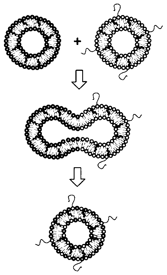
[0096] (3) Freeze the solution obtained above for 5 to 10 minutes at ultra-low temperature, thaw it by ultrasonication at room temperature for 30 to 60 minutes, and repeat the above freezing and thawing steps several times to obtain exosome-liposome complexes, liposome-exosome complexes body membrane fusion image 3 shown.
[0097] 2. Results analysis
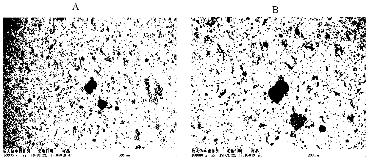
[0098] Depend on Figure 4 It can be seen that the nanoliposome-exosome complex is in the shape of spherical particles, and the particle ...
PUM
| Property | Measurement | Unit |
|---|---|---|
| Particle size | aaaaa | aaaaa |
| Particle size | aaaaa | aaaaa |
Abstract
Description
Claims
Application Information