Phage drug protein display system and application thereof

A protein display and phage technology, applied in the field of biomedicine, can solve the problems of increased secretion of immunosuppressive cytokines, cancer cell recognition, and reduced killing ability, achieving short amplification cycle, enhanced anti-tumor immune response, and simple operation Effect

Pending Publication Date: 2020-08-14
EAST CHINA UNIV OF SCI & TECH +1
View PDF5 Cites 1 Cited by
  • Summary
  • Abstract
  • Description
  • Claims
  • Application Information

AI Technical Summary

Problems solved by technology

However, with the development of tumors, the continuous anti-tumor immune response will lead to changes in the surface signaling molecules of tumor cells and immune cells, which will reduce the ability of immune cells to recognize and kill cancer cells, and at the same time increase the secretion of immunosuppressive cytokines, resulting in immune response. Inhibitory microenvironment, prompting tumor cells to evade immune mechanisms

Method used


Image

  • Phage drug protein display system and application thereof
  • Phage drug protein display system and application thereof
  • Phage drug protein display system and application thereof

Examples


Embodiment 1

[0066] Example 1. Nucleic acid vector construction

[0067] Use the NCBI database to search the human PD-1 gene sequence, select the coding gene of the extracellular segment of PD-1 protein (aa21-170), and optimize the sequence according to the codon preference of E. coli. Technology Co., Ltd. for gene synthesis.

[0068] Design the upstream and downstream primers P1 and P2 for gene amplification of the extracellular region of PD-1 protein:

[0069] P1: GCCATGGCCCAGGTGCAGCTGCCGGGTTGGTTCCTGGACTCTCCG

[0070] P2: CTGATATCTTTGGATCCAGCGGCCGCACCAACCAGGGTCTGGAACTGACCAG

[0071] Using the synthetic PD-1 gene as a template, the target gene was amplified by PCR using Super-Fidelity DNA polymerase. The PCR reaction system is: 25μL buffer, 20μL ddH 2 O, 1 μL dNTP, 1 μL primer P1, 1 μL primer P2, 1 μL template, 1 μL high-fidelity DNA polymerase. The PCR reaction program was: pre-denaturation at 95°C for 5 min, denaturation at 95°C for 20 s, annealing at 60°C for 30 s, extension at 72...

Embodiment 2

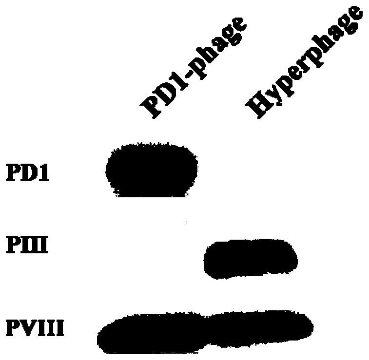
[0074] Example 2. Construction of PD-1 Display Phage

[0075] Inoculate 500 μL of the ER2738 strain carrying the pSEX81-PD-1 plasmid into 50 mL of 2YT medium (containing 100 μg / mL ampicillin and 100 mM glucose), and culture with shaking at 220 rpm at 37°C until OD600=0.5. Add M13KO7ΔPⅢ helper phage (the ratio of the number of Escherichia coli to the number of phages is 20:1) and mix well, put it in a 37°C incubator and culture it for 20 minutes, then place it in a shaker at 37°C at 220rpm and shake it for 45 minutes to make Escherichia coli obtain kanamycin resistance. Pour the bacterial solution into a centrifuge tube and centrifuge at 2000g for 10min at 4°C, discard the supernatant, add 50mL of fresh 2YT medium (containing 100μg / mL ampicillin, 50μg / mL kanamycin) to resuspend the bacteria, shake at 37°C Shake culture at 220rpm in the bed overnight.

[0076] Collect the bacterial solution and centrifuge at 2000g for 10min at 4°C, filter the supernatant with a 0.22μm sterile ...

Embodiment 3

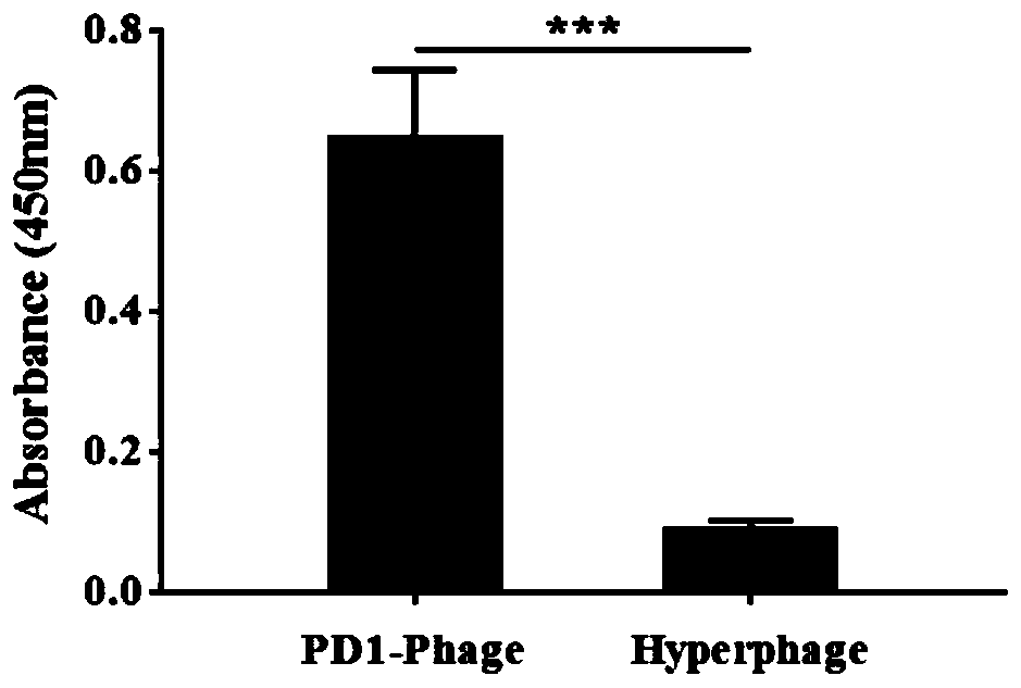
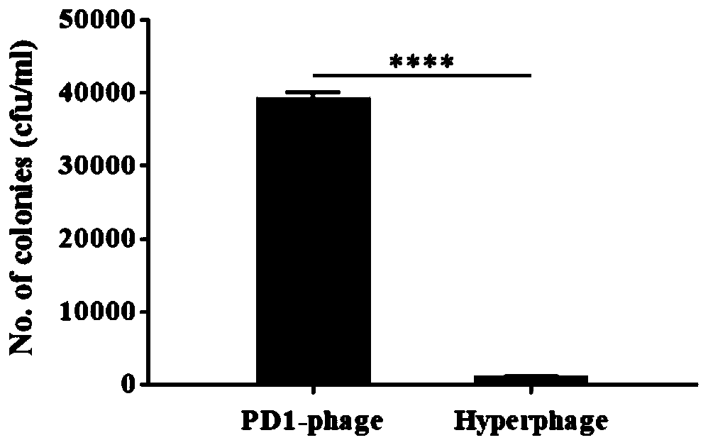
[0080] Example 3.PD-1 Displays Binding Specificity of Phage

[0081] with dilution buffer (100mM NaHCO 3 , pH 8.6) Dilute the PD-1 displaying phage and helper phage to 10 10 pfu / mL, 100 μL each was added to the wells of a 96-well microtiter plate, and incubated overnight in a 4°C refrigerator. TBST (PBS+0.5% Tween 20) was washed 5 times, and 200 μL of blocking solution (TBST containing 5% BSA) was added to each well to block at 37° C. for 1 hour. Wash 5 times with TBST, dilute rhPD-L1 to 1 μg / mL, add 100 μL to each well and incubate at 37 °C for 1 h. After washing 5 times with TBST, the mouse anti-PD-L1 monoclonal antibody was diluted with TBST at a ratio of 1:2000, and 100 μL was added to each well and incubated at 37°C for 1 h. After washing 5 times with TBST, the HRP-labeled goat anti-mouse secondary antibody was diluted with TBST at a ratio of 1:5000, and 100 μL was added to each well and incubated at 37°C for 1 h. Wash 5 times with TBST, add 100 μL TMB ELISA chromogen...


PUM


Abstract

The invention relates to the field of biological medicine, in particular to a phage drug protein display system and application thereof. The phage drug protein display system comprises engineering bacteria and a nucleic acid carrier, and the phage drug protein display system can generate phages for displaying drug proteins. According to the phage drug protein display system, a phage display technology is utilized, the phages with an immune checkpoint blocking function can be flexibly constructed according to specific requirements, and a new idea is provided for development of anti-tumor drugsand tumor treatment. The invention also discloses the application of the phage drug protein display system to enrich anti-tumor drug production and tumor treatment methods. According to the phage drugprotein display system provided by the invention, the high immunogenicity of the phage is combined with the immune checkpoint blocking function of the drug protein, so that the anti-tumor immune response is enhanced, and the better immune treatment effect is achieved.

Description

technical field [0001] The invention relates to the field of biomedicine, in particular to a phage drug protein display system and its application. Background technique [0002] Tumor is a major problem that threatens human health. It is a kind of disease characterized by abnormal cell proliferation. Traditional treatment methods include surgery, chemotherapy, radiotherapy, etc., but there are still many problems in these treatment methods, such as high toxicity to normal tissues and cells, poor targeting, and easy drug resistance, which prompts people to continuously research and develop new drugs. Tumor treatment methods. Studies have found that in the early stage of tumor cell colonization and growth, the immune system can effectively identify and destroy tumor cells to hinder the development of tumors. However, with the development of tumors, the continuous anti-tumor immune response will lead to changes in the surface signaling molecules of tumor cells and immune cell...

Claims


Application Information

Patent Timeline
Patent Type & Authority Applications(China)
IPC IPC(8): C12N15/70C12N1/21G01N33/68G01N33/574A61K35/76A61P37/04A61P35/00
CPCC12N15/70C07K14/70521G01N33/68G01N33/574A61K35/76A61P37/04A61P35/00
Inventor 叶邦策李虹锐
Owner EAST CHINA UNIV OF SCI & TECH
Why Eureka
Try Eureka