
A kind of nucleic acid nanostructure probe and its preparation method and application

A nucleic acid nano-probe technology, applied in biochemical equipment and methods, DNA preparation, chemical instruments and methods, etc., to achieve low detection limit, short synthesis time, and reduced loss efficiency

Active Publication Date: 2021-09-17
INST OF QUALITY STANDARD & TESTING TECH FOR AGRO PROD OF CAAS
View PDF1 Cites 0 Cited by
  • Summary
  • Abstract
  • Description
  • Claims
  • Application Information

AI Technical Summary

Problems solved by technology

[0004] Although there are some methods to detect APE1 and ROS in vivo and in vitro, it is difficult to detect APE1 and O in cells. 2 · - Simultaneous identification, localization and quantification

Method used


Image

  • A kind of nucleic acid nanostructure probe and its preparation method and application
  • A kind of nucleic acid nanostructure probe and its preparation method and application
  • A kind of nucleic acid nanostructure probe and its preparation method and application

Examples


Embodiment 1

[0083] In Vitro Recognition of APE1 Enzyme Using Nucleic Acid Nanoprobes

[0084] (1) Synthetic probes

[0085] (1) Dilute the seven DNA single strands P1, P2, P3, P4, P5-CY5, P6-BHQ3, and P7-HE to 25 μM with enzyme-free water, and vortex for 10 seconds to fully mix the single strands.

[0086] (2) To establish a 50 μL system, add 31 μL enzyme-free water and 5 μL 10×TAE buffer-Mg 2+ , 2 μL P1, 2 μL P2, 2 μL P3, 2 μL P4, 2 μL P5-CY5, 2 μL P6-BHQ3, 2 μL P7-HE; add the sample on the side wall of the centrifuge tube, after adding the sample, centrifuge and mix.

[0087] (3) The PCR annealing procedure was used to combine the seven DNA single strands into a three-dimensional nucleic acid nanostructure. The PCR setting program is as follows: 80°C, 10min; 4°C, 30min. Store at 4°C until use.

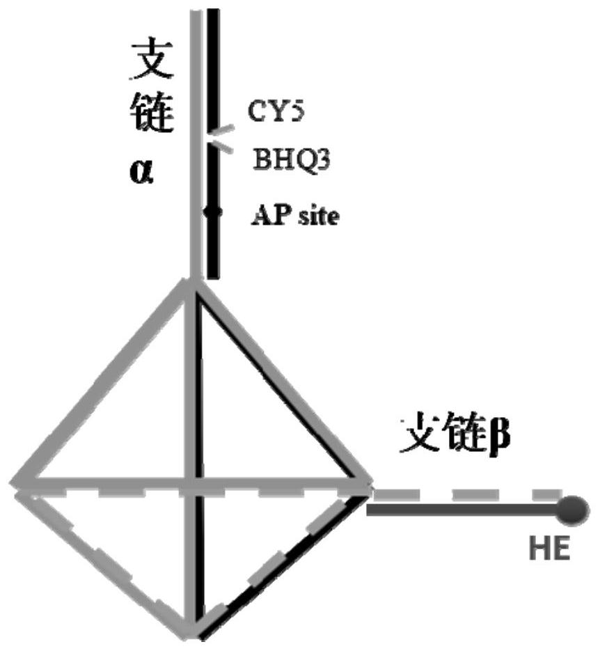
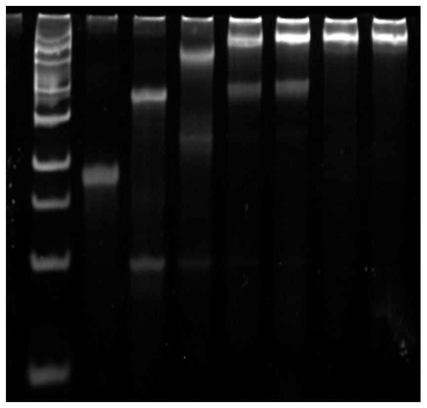
[0088] A schematic diagram of a nucleic acid nanostructure such as figure 1 Shown; Use polyacrylamide gel electrophoresis to the result that the probe synthesized by the embodiment of the p...

Embodiment 2

[0092] In vitro recognition of superoxide anion O using nucleic acid nanoprobes 2 · -

[0093] (1) Synthetic probes

[0094] Method is with embodiment 1 (one)

[0095] (2) In vitro identification and results

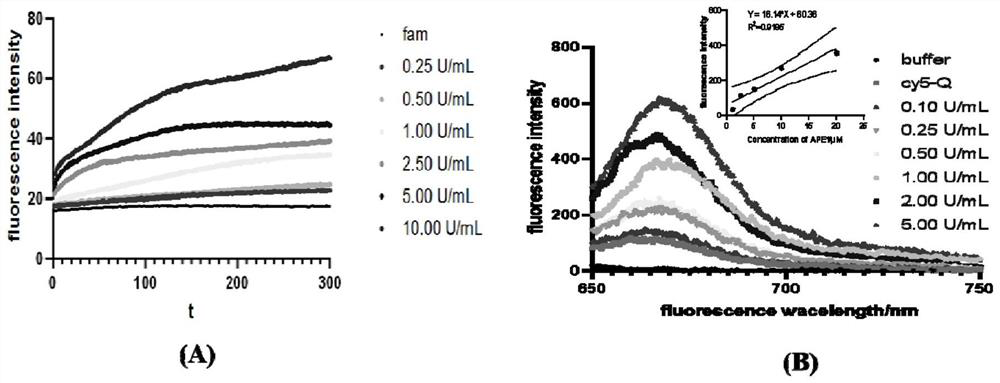
[0096] The synthesized probe was reacted with different concentrations of xanthine / xanthine oxidase in PBS buffer solution, and the fluorescence response intensity was between Figure 4 shown in . The experiment was performed in a fluorimeter. 1mol of xanthine and 1U of xanthine oxidase can generate 0.33mol of superoxide anion. The initial velocity of the fluorescence-concentration curve showed a good linear relationship between the probe and the concentration of superoxide anion in the range of 0.33-133.33mol / L. The detection limit was determined to be 0.33mol / L.

Embodiment 3

[0098] Application of nucleic acid nanoprobes to study APE1 expression and redox levels in cells exposed to PMA

[0099] PMA (phorbol ester) is a stimulator of reactive oxygen species. Cells exposed to PMA will lead to an increase in the content of reactive oxygen species and an imbalance in redox levels.

[0100] (1) Synthetic probes

[0101] Method is with embodiment 1 (one)

[0102] (2) Cell fluorescence experiment

[0103] (1) Cell receiving dish: Take out the tested cells from the constant temperature incubator, wash the cells with 1×PBS, draw 1ml of fresh complete medium with a pipette gun and repeatedly suck and blow the bottom of the culture flask, and mix the cells evenly. Pipette 1ml of the cell mixture into a 1.5ml EP tube and centrifuge at 1000rmp for 3min. Discard the supernatant after centrifugation, blow and mix the cells with fresh complete medium, pipette 200 μL of the uniform mixture into the groove of the confocal culture dish, dilute the cell suspension ...


PUM


Abstract

The invention discloses a nucleic acid nanostructure probe, its preparation method and application, and relates to the technical field of molecular detection. The nano-nucleic acid probes include: a. a nanomolecular cage with a tetrahedral structure composed of 4 main-chain DNA single strands; wherein the 2 main-chain DNA single-strands have branched chains α and branched chains extending out of the tetrahedral structure respectively. Chain β; b. Two non-main-chain DNA single strands combined with the branched chain α through complementary base pairing, one of which is modified with a fluorescent group, and the other is modified with an AP site and a quencher group; c. Another non-main-chain DNA single strand containing dihydroethidium combined with the branched-chain β through complementary base pairing. The invention also discloses the preparation method and application of the nucleic acid nanostructure. The nucleic acid nanoprobe of the present invention can simultaneously detect APE1 and O in cells 2 · ‑ , can be used for the simultaneous determination of nucleic acid repair enzymes and reactive oxygen species. Through the confocal imager, the target detection substances APE1 and O in cells can be detected 2 · ‑ Check the spatial distribution.

Description

technical field [0001] The invention relates to the technical field of molecular detection, in particular to a nucleic acid nanostructure probe and its preparation method and application. Background technique [0002] The base excision repair pathway (base excision repair, BER) in cells is a key step in responding to oxidative stimuli, and has special significance for maintaining the stability of the cell genome. Apurinic / apyrimidinic (AP) endonuclease 1 (APE1), a multifunctional protein, is a key enzyme involved in the recognition and processing of AP sites in the BER pathway, while maintaining the balance of the intracellular environment makes an important impact. Under endogenous or exogenous stimuli, the redox system of cells will be out of balance, and APE1 can directly or indirectly regulate the redox state of cells, thereby helping to maintain the homeostasis of cells. APE1 has redox activity and base repair activity, so it is also called redox factor-1, Ref-1. Fur...

Claims


Application Information

Patent Timeline
Patent Type & Authority Patents(China)
IPC IPC(8): C12Q1/527C12N15/11C12N15/10C09K11/06G01N21/64
CPCC09K11/06C09K2211/1029C12Q1/527G01N21/6428
Inventor 钱永忠潘烨灿苏昕翁瑞邱静
Owner INST OF QUALITY STANDARD & TESTING TECH FOR AGRO PROD OF CAAS
Learn More