
Template for tissue engineering material and tissue engineering material

A technology of tissue engineering and templates, applied in medical science, blood vessels, prostheses, etc., can solve the problems of coagulation matrix adhesion, easy sliding, and exposed skeleton fibers

Pending Publication Date: 2020-09-25
领博生物科技(杭州)有限公司
View PDF7 Cites 0 Cited by
  • Summary
  • Abstract
  • Description
  • Claims
  • Application Information

AI Technical Summary

Problems solved by technology

However, in this process, the uniformity of the parallel spacing of the fibers in each layer of the polymer backbone is poor, resulting in uneven pore diameters of the backbone, resulting in a low yield of blood vessels
In addition, the fibrous structure of the skeleton is also prone to structural damage due to subcutaneous implantation operations or in vitro cell culture, reducing the yield of blood vessels
And on the basis of the previous research, we also found that the outer surface of the prepared polymer skeleton-reinforced tissue engineered blood vessel is relatively smooth, and it is easy to slip after implantation in the body, and the integration rate with the surrounding tissues of the implantation site needs to be improved. Some of the skeleton fibers are exposed in the lumen of polymer skeleton-reinforced tissue engineered blood vessels, which will cause a certain degree of coagulation matrix adhesion

Method used


Image

  • Template for tissue engineering material and tissue engineering material
  • Template for tissue engineering material and tissue engineering material
  • Template for tissue engineering material and tissue engineering material

Examples


Embodiment 1

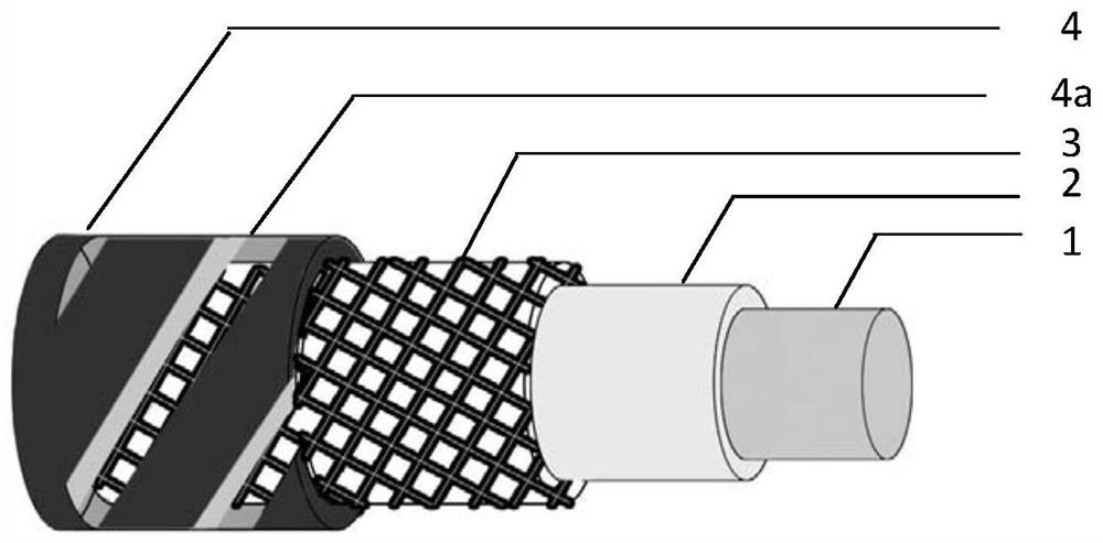
[0051] The tissue engineering material template provided in this embodiment is a tissue engineering blood vessel template, which includes a support body, that is, an inner core, an anti-exposure layer, a reticular fiber skeleton layer, and a protective layer, that is, an overcoat from the inside to the outside; the protective layer is provided with Hollow structure; the protective layer is arranged outside the mesh fiber skeleton layer, and the inner diameter of the protective layer matches the outer diameter of the mesh fiber skeleton layer.

[0052] The template preparation process includes the following steps:

[0053] 1. Select a silicone tube as the support, with an outer diameter of 2 mm and an inner diameter of 1 mm; the outer diameter of the support determines the inner diameter of the prepared tissue engineering blood vessel.

[0054] 2. Using PDS: gelatin = 1:2 (mass ratio) as the raw material, the biodegradable layer was prepared on the surface of the silicone tube ...

Embodiment 2

[0066] The template preparation process includes the following steps:

[0067] 1. A polyurethane tube with an outer diameter of 4 mm and an inner diameter of 2 mm is used as a support body, and the outer diameter of the support body determines the inner diameter of the prepared tissue engineering blood vessel.

[0068] 2. Using PLGA and PEO as raw materials, high-voltage electrospinning technology is used to prepare an anti-exposure layer on the surface of the polyurethane tube. The specific method is: insert a stainless steel rod with a suitable diameter into the above polyurethane tube as a receiving rod, and use chloroform: methanol =5:1 (volume ratio) as a solvent, PLGA:PEO=1:1 (mass ratio) as a solute, prepare an electrospinning solution with a concentration of 0.28g / ml, use a 20G needle, and set a flow rate of 8mL / h. A DC voltage of 16kV is applied to the spinning needle, and a grounded stainless steel plate is used as the conductive receiving plate. The receiving rod is...

Embodiment 3

[0076] 1. A silicone tube with an outer diameter of 5 mm and an inner diameter of 3 mm is used as a support body, and the outer diameter of the support body determines the inner diameter of the prepared tissue engineering blood vessel.

[0077] 2. Using P(3HB-co-4HB) and PEO as raw materials, high-voltage electrospinning technology is used to prepare an anti-exposure layer on the surface of the silicone tube. The specific method is: insert a stainless steel rod with a suitable diameter into the above-mentioned silicone tube as a receiving Rod, with chloroform: methanol = 5:1 (volume ratio) as solvent, P(3HB-co-4HB): PEO = 1:0.5 (mass ratio) as solute, and prepare electrospinning with a concentration of 0.3g / ml Liquid, using a 19G needle, set the flow rate to 8mL / h, apply a DC voltage of 14kV to the needle, use a grounded stainless steel plate as the conductive receiving plate, place the silicone tube between the conductive receiving plate and the needle, and the distance from t...


PUM

PropertyMeasurementUnit
Thicknessaaaaaaaaaa
Widthaaaaaaaaaa
Thicknessaaaaaaaaaa

Abstract

The invention provides a template for a tissue engineering material. The template comprises a support body, an anti-exposure layer, a reticular fiber skeleton layer and a protective layer which are sequentially arranged, wherein a hollow structure is arranged on the protective layer. Compared with the prior art, when the template provided by the invention is used for preparing the tissue engineering material, the anti-exposure layer is degraded to form a gap, and finally, the surface of an inner cavity of a reticular fiber skeleton can be coated with an acellular matrix material, so that fiberexposure is reduced, and adhesion of blood coagulation components is reduced; the reticular fiber skeleton can provide good mechanical properties, so that the tissue engineering material has good kink resistance, resilience, burst resistance, puncture closeness and suturing property; meanwhile, the protective layer can protect the structure of the reticular fiber skeleton layer, so that the skeleton structure is prevented from being damaged when the material is cultured and prepared; and due to the arrangement of the hollow structure on the protective layer, the prepared tissue engineering material can be provided with an external ridge, the suture strength, burst pressure and kink resistance of the blood vessels are enhanced, and the integration with the surrounding tissues is accelerated.

Description

technical field [0001] The invention belongs to the technical field of tissue engineering, and in particular relates to a template for tissue engineering materials and tissue engineering materials. Background technique [0002] Cardiovascular disease is the disease with the highest fatality rate in the world. The occurrence of this disease is often due to vascular stenosis or blockage, which leads to reduced blood flow and lack of nutrients, which damages tissues or organs. It usually manifests as coronary heart disease, cerebrovascular disease, peripheral arterial disease. According to the prediction of the World Health Organization, by 2030, the number of people dying of cardiovascular-related diseases in the world will increase to 23.3 million each year. Vascular transplantation is still a routine method for the treatment of such diseases. At present, the collection and use of patients' autologous blood vessels (such as great saphenous vein, internal thoracic artery on b...

Claims


Application Information

Patent Timeline
IPC IPC(8): A61F2/06A61L27/18A61L27/20A61L27/22A61L27/24A61L27/36A61L27/50A61L27/58
CPCA61F2/06A61L27/3604A61L27/18A61L27/222A61L27/24A61L27/20A61L27/225A61L27/507A61L27/58Y02W90/10
Inventor 许鹏赟徐霁季旭东
Owner 领博生物科技(杭州)有限公司
Learn More