Separation and preparation methods of human amniotic epithelial stem cells

A separation method and stem cell technology, applied in the field of preparation, serum-free culture method and its products, and separation of human amniotic epithelial stem cells, can solve the problems of animal-derived pathogen contamination, foreign protein rejection, amniotic epithelial cell destruction, etc.

Inactive Publication Date: 2020-10-20
SHANGHAI ICELL BIOTECH +1
View PDF8 Cites 1 Cited by
  • Summary
  • Abstract
  • Description
  • Claims
  • Application Information

AI Technical Summary

Problems solved by technology

[0011] In order to solve the technical problems of damage to amniotic epithelial cells caused by the existing method for separating human amniotic membrane epithelial stem cells, and the contamination of animal-derived pathogens caused by residual animal serum in the cells and the rejec

Method used


Image

  • Separation and preparation methods of human amniotic epithelial stem cells
  • Separation and preparation methods of human amniotic epithelial stem cells
  • Separation and preparation methods of human amniotic epithelial stem cells

Examples


Embodiment 1

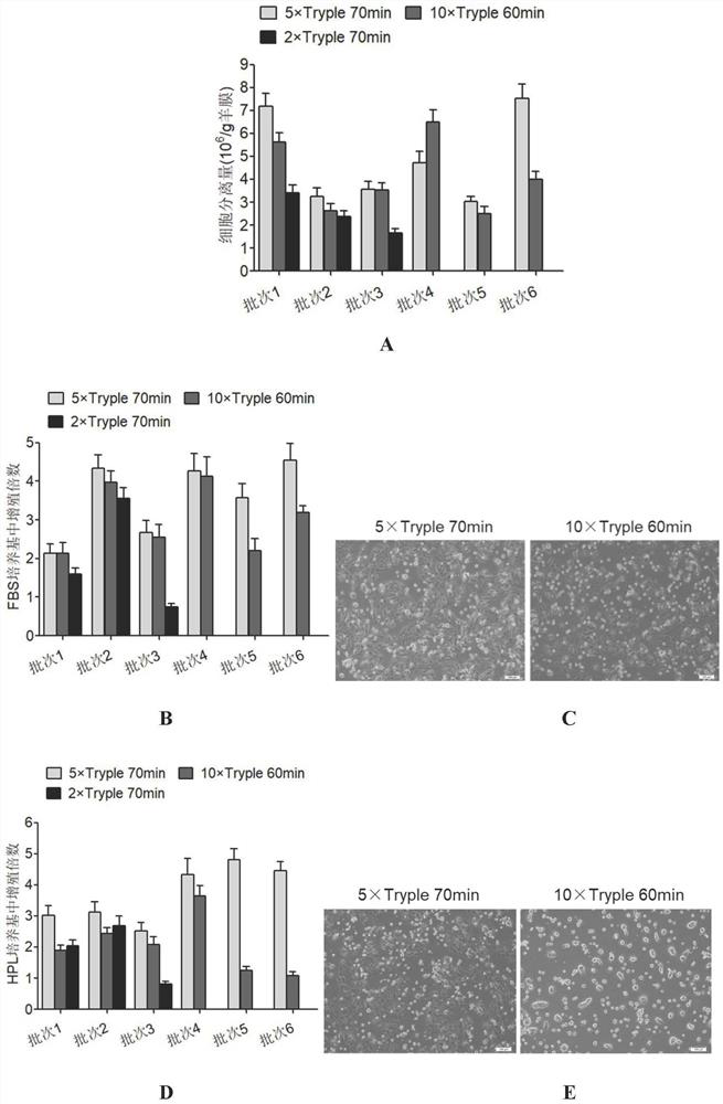
[0063] Example 1 Enzyme Digestion and Separation Conditions for Human Amniotic Membrane Epithelial Stem Cells

[0064] With the informed consent of the puerpera, the placental amniotic membranes of healthy fetuses who underwent cesarean section before 39 weeks of gestation were collected. After testing, the mother is in good health and has no acute or chronic infectious diseases, family genetic diseases, hepatitis B virus, hepatitis C virus, HIV, human T cell-tropic virus, herpes virus, cytomegalovirus, Treponema pallidum and mycoplasma and other pathogens , The collection of amniotic membrane was carried out under sterile conditions.

[0065] After the amniotic membrane is collected, it is placed in the amniotic membrane preservation solution, transported at 4°C, and arrives at the GMP cell preparation workshop within 12 hours for digestion and separation of human amniotic membrane epithelial stem cells.

[0066] The amniotic membrane was taken out from the preservation solu...

Embodiment 2

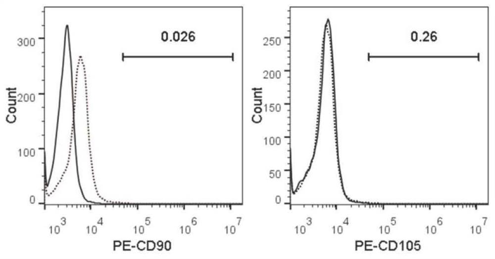
[0072] Embodiment 2 Freshly isolated human amniotic membrane epithelial stem cells (Pre-P0) phenotype detection

[0073] Pre-P0 cells were separated by trypsinization, and the cells were centrifuged and washed twice with FACS buffer (PBS containing 2% FBS), and the cells were resuspended in FACS buffer, and the cell concentration was adjusted to 1×10 6 / 100μL, split into several tubes, add CD90, CD105 and their respective isotype control antibodies to the cells in each tube, incubate at 4°C in the dark for 30min, centrifuge and wash the cells twice with FACS buffer, resuspend in 500μL FACS buffer Cells, immediately tested on the machine.

[0074] see figure 2 , the results showed that the Pre-P0 cells separated by trypsin digestion did not express two classic mesenchymal stem cell markers, CD90 and CD105, indicating that the isolated human amniotic epithelial stem cells had no mesenchymal stem cell contamination and were of high purity.

Embodiment 3

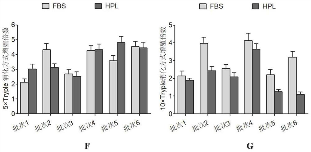
[0075] Example 3 Screening of human amniotic epithelial stem cells in serum-free medium

[0076] The Pre-P0 cells were resuspended in 6 kinds of media, respectively, FBS (DMEM / F-12, 10% concentration of fetal bovine serum, 1mM sodium pyruvate, 1mM non-essential amino acids, 2mM L-glutamine and 10 ng / mL human epidermal growth factor), HPL (DMEM / F-12, 10% platelet lysate, 1mM sodium pyruvate, 1mM non-essential amino acids, 2mM L-glutamine and 10ng / mL human epidermal growth factor), B27 (DMEM / F-12, add 2% volume of Gibco B27supplement, 1mM sodium pyruvate, 1mM non-essential amino acids, 2mM L-glutamine and 10ng / mL human epidermal growth factor), CTS B27 (DMEM / F-12 , add 2% volume of Gibco's CTSB27 supplement, 1mM sodium pyruvate, 1mM non-essential amino acids, 2mML-glutamine and 10ng / mL human epidermal growth factor), N2 (DMEM / F-12, add 1% volume of Gibco company N2supplement, 1mM sodium pyruvate, 1mM non-essential amino acids, 2mM L-glutamine and 10ng / mL human epidermal growth...


PUM


Abstract

The invention discloses separation and preparation methods of human amniotic epithelial stem cells (hAESCs) and the hAESCs. The separation method comprises the following steps of (1) pretreating amniotic tissue with recombinant trypsin, and removing the recombinant trypsin to obtain pretreated amniotic tissue; (2) digesting the pretreated amniotic tissue obtained in the step (1) by using the recombinant trypsin, and collecting digestive juice; (3) adding a digestive terminator into the digestive juice obtained in the step (2), performing filtration and centrifugation on the obtained digestivejuice, discarding a supernatant, and collecting precipitated cells; and (4) inoculating the cells obtained in the step (3) into a serum-free and animal-source-free culture medium system, and performing culture and amplification to obtain the high-purity hAESCs. The hAESCs have the advantages of being rapid to separate, easy and convenient to operate, low in cost and the like, and the obtained hAESCs are directly used for scientific research and clinical treatment and have wide application prospects.

Description

technical field [0001] The invention belongs to the field of biological technology, and in particular relates to a method for separating, preparing, and culturing human amniotic membrane epithelial stem cells without serum and a product thereof. Background technique [0002] The amniotic membrane is the innermost layer of the placenta, located on the surface of the fetal chorion. It is an early product of embryonic development that is closely connected with the developing fetus, and is also an important tissue for material exchange between the mother and the fetus. The amniotic membrane is a translucent membrane with toughness, without nerves, blood vessels and lymphatic vessels, which wraps the amniotic fluid and fetus and provides an environment for embryonic growth and development. Its thickness is only 0.02 ~ 0.5mm, composed of epithelial layer, basal layer and stroma (connective tissue layer). Between the stromal layer of the amniotic membrane and the chorion there is ...

Claims


Application Information

Patent Timeline
IPC IPC(8): C12N5/074
CPCC12N5/0607C12N2509/00C12N2500/90
Inventor 袁惟芯李宁王莉邵小燕黄晓军赵翔宇
Owner SHANGHAI ICELL BIOTECH
Why Eureka
Try Eureka