In-vitro induced immunosuppressive myeloid suppressive cell as well as preparation and application thereof

A technique of inhibiting cells and immunosuppression, applied in the field of cell biology and clinical application, which can solve the problems of unstable effect and lack of in vitro induction technology

Active Publication Date: 2020-10-23
THE SECOND XIANGYA HOSPITAL OF CENT SOUTH UNIV
View PDF23 Cites 2 Cited by
  • Summary
  • Abstract
  • Description
  • Claims
  • Application Information

AI Technical Summary

Problems solved by technology

[0006] In order to solve the technical problems that the existing immunosuppressive myeloid suppressor cells are mainly extracted in vivo, the effect is unstable, and the in vitro induction technology is lacking, the first purpose of the present invention is to provide a preparation method for in vitro induction of immunosuppressive myeloid suppressor cells , aiming to achieve high yield in vitro directional induction preparation of immunosuppressive function MDSC (ex vivo generated MDSC by cytokine-induced differentiation of bone marrow cells, evMDSC)

Method used


Image

  • In-vitro induced immunosuppressive myeloid suppressive cell as well as preparation and application thereof
  • In-vitro induced immunosuppressive myeloid suppressive cell as well as preparation and application thereof
  • In-vitro induced immunosuppressive myeloid suppressive cell as well as preparation and application thereof

Examples


Embodiment 1

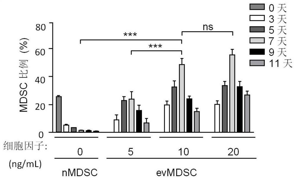
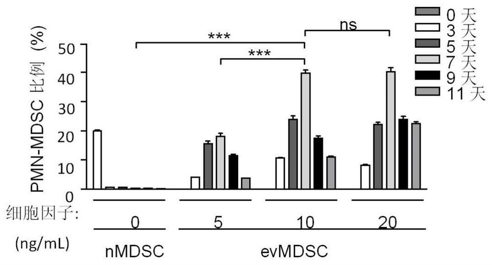
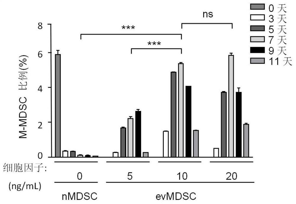
[0055] Establishment of Immunosuppressive Functional MDSC System in Vitro

[0056] (1) Obtaining mouse bone marrow cells: under sterile conditions, 6-8 week old male BALB / c mice were sacrificed, and bilateral tibia and fibula were taken, the medullary cavity was washed with PBS, and the cells were collected in a centrifuge tube to obtain nMDSC cells.

[0057] (2) Preparation of culture medium: prepare basal medium according to 10% fetal bovine serum, 90% 1640 medium, 1% penicillin-streptomycin double antibody, add different concentrations of cytokines GM-CSF and IL-6 (0 , 5, 10, 20ng / ml; the ratio of GM-CSF and IL-6 is 1:1).

[0058] (3) Cell inoculation and culture: the cells were divided into 5x10 5 cells / well were seeded in a 24-well plate, and medium containing different concentrations of cytokines was added, and the cells were incubated at 37°C, 5% CO 2 and saturated humidity for different days (0, 3, 5, 7, 9, 11 days) respectively, and half the volume of medium was str...

Embodiment 2

[0063] CD4 + T cell proliferation inhibition assay

[0064] (1) Obtain mouse CD4 + T cells: Dissect and dissociate the spleen of 6-8 week male Balb / c mice in a sterile environment, collect the liquid after grinding, filter it through a 200-mesh nylon mesh and collect it in a centrifuge tube, add red blood cell lysate 3 times the volume of the cell, Labeled with magnetic beads and combined with CD4 monoclonal antibody, CD4 was obtained by magnetic column separation + T. After sorting, the cells were tested for separation purity with a flow cytometer.

[0065] (2) CD4 + T cell CFSE staining: CD4 + T cell suspension was washed twice with PBS at room temperature and resuspended to 5-10x10 6 cells / mL, add 1uM CFSE to the final concentration, mix and incubate at room temperature for 10min in the dark, add 4-5 times the volume of complete medium to stop incubation, incubate on ice for 5min, wash with complete medium for 3 times, add 0.4μg / mL anti -mouse CD28 monoclonal antibod...

Embodiment 3

[0070] Effect of MDSC depletion in vivo on pathological neovascularization after penetrating keratoplasty

[0071](1) Establish a penetrating corneal transplantation mouse model: Pure-line 6-8 week male Balb / c mice are used as recipients, and C57BL / 6 mice are used as donors for corneal transplantation, which is called allogeneic corneal transplantation (Allograft, referred to as Allo). The donor and recipient are both BALB / c, which is called allogeneic corneal transplantation (Isograft, Iso for short). The recipient cornea was sutured in advance to induce the neovascularization state of the implant bed, and the corneal transplantation was performed one week later: the graft was taken with a diameter of 2.25 mm, the diameter of the implant bed was 2.0 mm, and the graft was sutured watertightly on the implant bed with continuous suture of 11 / 0 silk thread. The eyelids were closed with 8 / 0 silk sutures. Eyelid sutures were removed on the third day after surgery, and corneal sut...


PUM


Abstract

The invention relates to a preparation and acquisition method of in-vitro induced immunosuppressive myeloid suppressive cells, and belongs to the field of cytobiology and clinical application. The invention specifically discloses an immunosuppressive myeloid suppressor cell (MDSCs) which is derived from a healthy donation individual and generates immunosuppressive activity through in-vitro drug induction stimulation, and a preparation and purification method of the immunosuppressive myeloid suppressor cell. The invention finds that in vitro induced immunosuppressive myeloid suppressor cells can effectively inhibit CD4 + T cell proliferation effect in vitro; by means of the application, the biological preparation for effectively treating rejection after corneal transplantation and effectively inhibiting pathological angiogenesis can be prepared.

Description

technical field [0001] The invention relates to a method for preparing and obtaining immunosuppressive myeloid suppressor cells induced in vitro, and belongs to the field of cell biology and clinical application. Background technique [0002] Myeloid-derived suppressor cells (Myeloid-Derived Suppressor Cells, MDSC) are myeloid progenitor cells derived from bone marrow with immature morphology, which can differentiate downstream into mature granulocytes, macrophages or dendritic cells under certain circumstances. In mice, MDSC characteristically co-express surface molecules myeloid differentiation antigen (Myeloid-cell Lineage Differentiation Antigen-1, Gr-1) and CD11b (ie αM-integrin). According to the expression of Gr-1 subgroup Ly6C and Ly6G in MDSC, it can be roughly divided into granular MDSC (CD11b + Gr-1 + Ly6G high Ly6C - , PMN-MDSC) and mononuclear MDSC (CD11b + Gr-1 + Ly6G - Ly6C high , M-MDSC) two groups (VegliaF, Perego M, GabrilovichD. Myeloid-derived sup...

Claims


Application Information

Patent Timeline
Patent Type & Authority Applications(China)
IPC IPC(8): C12N5/077A61K35/15A61P37/06
CPCC12N5/0669A61K35/15A61P37/06C12N2501/2306C12N2501/22
Inventor 何彦凌林周也荻任悦容陈百华
Owner THE SECOND XIANGYA HOSPITAL OF CENT SOUTH UNIV
Why Eureka
Try Eureka