Bionic three-dimensional scaffold for promoting bone defect repair and preparation method thereof

A technology for three-dimensional scaffolds and bone defects, applied in the field of bionic three-dimensional scaffolds and their preparation, can solve the problems of difficult control of inter-lamellar spacing, unfavorable adhesion and proliferation of bone marrow penetrating cells, preventing displacement or even scattering, facilitating cell adhesion and proliferation, Size controllable effect

Active Publication Date: 2021-01-26
XUZHOU MEDICAL UNIV
View PDF6 Cites 2 Cited by
  • Summary
  • Abstract
  • Description
  • Claims
  • Application Information

AI Technical Summary

Problems solved by technology

However, due to the limitations of the wrapping process, it is difficult to control the distance between the layers, which is not conducive to the penetration of the bone marrow and the adhesion and proliferation of cells.

Method used


Image

  • Bionic three-dimensional scaffold for promoting bone defect repair and preparation method thereof
  • Bionic three-dimensional scaffold for promoting bone defect repair and preparation method thereof
  • Bionic three-dimensional scaffold for promoting bone defect repair and preparation method thereof

Examples


Embodiment 1

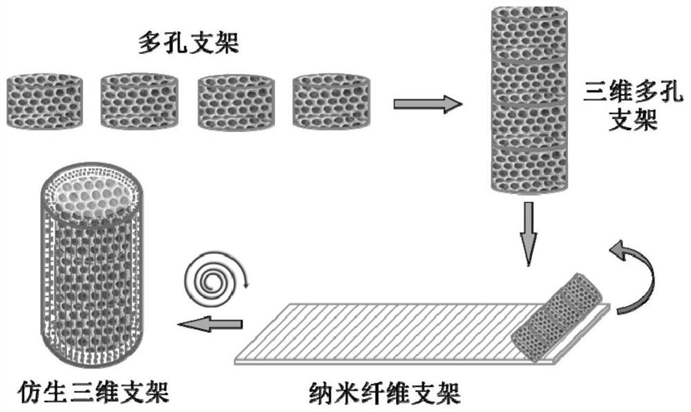
[0026] 1. Obtaining of PLGA electrospun nanofibers: 3g PLGA was dissolved in 100ml chloroform (CHCl 3 ) and N,N-dimethylformamide (DMF) mixed solution (volume ratio 3:1) to obtain a transparent solution, put it into a syringe, the syringe needle is flat, the inner diameter is 0.5mm, and the syringe is fixed on On the propulsion pump, the propulsion speed is set to 1.5ml / h, the positive pole of the high-voltage DC power supply is connected to the syringe needle, the negative pole is connected to the plate or roller collector, the voltage is 15kV, the collection distance is 25cm, the time is 1h, and the PLGA static electricity is obtained by vacuum drying at 30°C for 24h. Spun nanofibrous membrane (attached figure 2 ). (For specific methods, please refer to Colloids and Surfaces B: Biointerfaces2018, 167, 550-559)

[0027] 2. The acquisition of the PLGA porous support: collect gelatin microspheres with a microfluidic device, and the microfluidic conditions are as follows: the...

Embodiment 2

[0030] It is basically the same as Example 1, except that the outer PLGA electrospun membrane is replaced by a PHBV electrospun membrane. 0.9g PHBV and 0.1g polyoxyethylene (PEO) were dissolved in 50ml trifluoroethanol (TFE); the solution was added to the syringe, the needle of the syringe was flat and the inner diameter was 0.5mm, the syringe was fixed on the propulsion pump, and the propulsion speed was set to 5ml / h, the positive electrode of the high-voltage DC power supply is connected to the needle of the syringe, and the negative electrode is received by the collector. The voltage is 12kV, and the collection distance is 25cm. After 2h, the obtained PHBV nanofiber membrane is removed, and vacuum-dried at 30°C for 24h to obtain PHBV electrospun fibers membrane. (The specific method can refer to the patent ZL201110037596.9)

Embodiment 3

[0032] It is basically the same as Example 1, except that the outer layer of PLGA nanofibers is doped with nano-sized hydroxyapatite (nHA). Dissolve 3 g PLGA in 75 ml CHCl 3 0.3g nHA was ultrasonically dispersed in DMF, and then the two were mixed and stirred evenly as an electrospinning solution, and PLGA / nHA nanofibers were obtained under the same conditions as in Example 1. (For specific methods, please refer to ACS Appl.Mater.Interfaces 2013,5,319-330)


PUM

PropertyMeasurementUnit
thicknessaaaaaaaaaa
diameteraaaaaaaaaa
diameteraaaaaaaaaa

Abstract

The invention discloses a bionic three-dimensional scaffold for promoting bone defect repair and a preparation method thereof. The scaffold is formed by compounding two units, wherein one unit is an inner-layer scaffold, is a three-dimensional porous scaffold with an inverse opal structure and is of a columnar structure, and a diameter and a height of the inner-layer scaffold can be adjusted according to a diameter and a defect part of a bone; and the other unit is an outer-layer scaffold, is an electrostatic spinning nanofiber film and wraps an outer part of the inner-layer scaffold, and a size of the film can be cut as required. The bionic three-dimensional scaffold simulates bone structure, a nanofiber multilayer structure on the outer layer simulates bone cortex, and the porous scaffold on the inner layer can allow bone marrow to pass and provide a suitable place for cell adhesion, and the bionic three-dimensional scaffold can promote bone repair and functional reconstruction. By controlling a size of the inner-layer scaffold and a size of the outer-layer film, a size of the bionic three-dimensional scaffold can be flexibly adjusted to meet needs of bone defects of different lengths.

Description

technical field [0001] The invention relates to a bionic three-dimensional support for promoting bone defect repair and a preparation method thereof, belonging to the technical field of biological tissue engineering. Background technique [0002] Bone has a certain ability to regenerate and repair, but in many cases, it cannot be repaired solely by its own regenerative ability, such as nonunion caused by external factors, bone defect after infection or bone tumor resection, orthopedic surgery, etc. In recent years, bone trauma caused by frequent natural disasters, bone trauma caused by a sharp increase in vehicle accidents, and bone loss caused by increasingly serious aging have made the demand for bone repair increasingly urgent. Bone transplantation has become the second tissue transplantation in the world, with as many as 3.7 million people undergoing transplantation to treat bone defects every year. [0003] Autologous bone transplantation is regarded as the gold standa...

Claims


Application Information

Patent Timeline
Patent Type & Authority Applications(China)
IPC IPC(8): A61L27/12A61L27/18A61L27/56A61L27/58A61F2/28
CPCA61L27/58A61L27/56A61L27/18A61L27/12A61F2/28A61F2/2846A61L2400/08A61L2430/02A61F2002/2835C08L67/04
Inventor 吕兰欣张晓峰张静逸刘敬芝沈红先胡书群燕宪亮
Owner XUZHOU MEDICAL UNIV
Why Eureka
Try Eureka