
Novel coronavirus antibody detection kit as well as preparation method and application thereof

A coronavirus and kit technology, applied in the field of cell biology and immunoassay, can solve the problems of cumbersome operation, cumbersome steps and high sensitivity, and achieve the effects of high detection efficiency, simple operation and good repeatability

Active Publication Date: 2021-02-19
ACROBIOSYSTEMS INC
View PDF4 Cites 1 Cited by
  • Summary
  • Abstract
  • Description
  • Claims
  • Application Information

AI Technical Summary

Problems solved by technology

The ELISA method is simple and common. This method has relatively high sensitivity and is less difficult to standardize the carrier. However, the detection speed is slow, easy to contaminate, and the steps are cumbersome.
The colloidal gold method is characterized by no need for special treatment of specimens, simple operation, short time-consuming, no detection equipment, and low restrictions on personnel and places for detection technology. It can be widely used in primary medical units and on-site detection. Determination can only be a qualitative test, not quantitative, nor can it be compared with antibody titers
S-Flow flow detection is based on the 293T cell line overexpressing the S protein, which is limited by the cell culture conditions and cell state, and can only detect the antibody of the S protein, and cannot take into account the detection of the antibody of the N protein at the same time
The luciferase immunoprecipitation method is to couple luciferase to S protein or N protein, after the antibody is bound to the protein, and then the luciferase-S protein or N protein-antibody The complex is precipitated, and the luciferase substrate chromogenic kit is used for chromogenic detection. This method is cumbersome to operate, and the detection sensitivity is lower than that of ELISA and S-Flow

Method used


Image

  • Novel coronavirus antibody detection kit as well as preparation method and application thereof
  • Novel coronavirus antibody detection kit as well as preparation method and application thereof
  • Novel coronavirus antibody detection kit as well as preparation method and application thereof

Examples


Embodiment 1

[0057] Example 1. Construction of novel coronavirus antibody detection kit

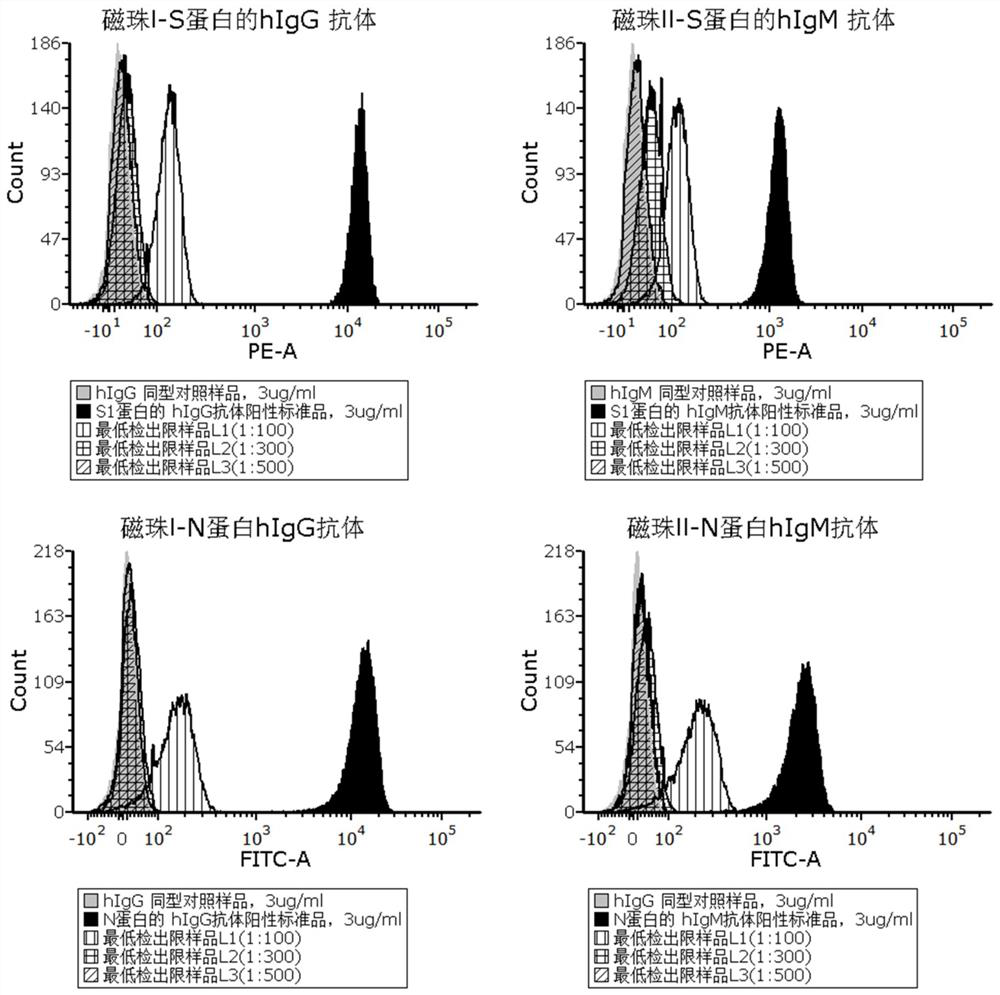
[0058] The magnetic bead fluorescent immunoassay kit for novel coronavirus antibody provided in this example has a specification of 100TEST and includes the following components:

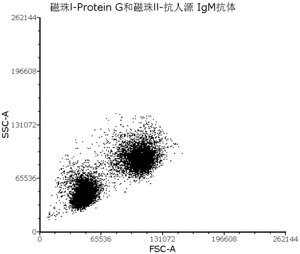
[0059] 1. Magnetic beads Ⅰ-Protein G, the specification is 1ml; magnetic beads I have a particle size of 2.8 μm, and are stored in a buffer, the buffer is 0.01M phosphate buffer containing 0.03% sodium azide, pH 7.4, the amount used 10μl / sample;

[0060]2. Magnetic beads II-anti-human IgM antibody, the specification is 1ml; the particle size of magnetic beads II is 10μm, and it is stored in buffer solution, the buffer solution is 0.01M phosphate buffer containing 0.03% sodium azide, pH7.4, The amount used is 10 μl / sample;

[0061] 3. PE-labeled novel coronavirus S1 protein, in the form of lyophilized powder, 1 vial, with a specification of 10 μg;

[0062] 4. FITC-labeled novel coronavirus N protein, in the form of freeze...

Embodiment 2

[0071] Embodiment 2, kit operation method

[0072] When the kit of Example 1 is used, the operation method is as follows:

[0073] 1. Take the PE-labeled novel coronavirus S1 protein, add 100 μl sterile ultrapure deionized water to reconstitute, the concentration is 100 μg / ml, use the flow detection buffer to prepare the working solution with a concentration of 2 μg / ml, and set aside.

[0074] 2. Take FITC-labeled novel coronavirus N protein, add 100 μl sterile ultrapure deionized water to reconstitute, the concentration is 50 μg / ml, use the flow detection buffer to prepare a working solution with a concentration of 1 μg / ml, and set aside.

[0075] 3. Add 100 μl of sterile ultrapure deionized water to reconstitute the antibody positive standard and antibody isotype control sample, the concentration is 100 μg / ml, and then dilute a series of concentration gradients with flow detection buffer, the recommended concentration is 0.003 μg / ml ml, 0.03μg / ml, 0.3μg / ml and 3μg / ml, for l...

Embodiment 3

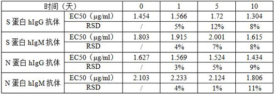
[0083] Embodiment 3, accelerated stability verification

[0084] Store the kit in Example 2 at 37°C for 0, 1, 5, and 10 days, and then detect the positive standard of the new coronavirus antibody. Each sample has 8 concentration gradients. Statistical analysis is performed on the detection data. The acceptance criteria are: compared with the control group (0 hour group), EC 50 The RSD value of the value is ≤20.0%. The measurement results at different times are shown in Table 1. Compared with the control group, the RSDs of EC50 values ​​were all less than 20%. The kits can be stored at -20°C for at least 12 months.

[0085] Table 1 The results of the accelerated stability test of the kit

[0086]


PUM


Abstract

The invention provides a novel coronavirus antibody detection kit as well as a preparation method and application thereof. The kit comprises an IgG binding molecule, an anti-IgM antibody, fluorescently-labeled novel coronavirus S1 protein, fluorescently-labeled novel coronavirus N protein, an hIgG antibody positive standard substance of the S1 protein, an hIgG antibody positive standard substanceof the N protein, an hIgM antibody positive standard substance of the S1 protein, an hIgM antibody positive standard substance of the N protein, a negative control hIgG antibody sample and a negativecontrol hIgM antibody sample, wherein the IgG binding molecule and the anti-IgM antibody are loaded on nanoparticles with different particle sizes. The kit disclosed by the invention is used for detecting novel coronavirus antibodies, can be used for quickly detecting novel coronavirus neutralizing antibodies in serum within 12 h, and has the advantages of small dosage of a to-be-detected sample,good specificity, high sensitivity, good repeatability, simplicity in operation, low laboratory requirement and high safety.

Description

technical field [0001] The present invention relates to a serological method for detecting novel coronavirus antibodies, in particular to a fluorescent immunoassay kit for detecting novel coronavirus IgG and IgM antibodies and its preparation method and application, belonging to cell biology and The field of immunoassay technology. Background technique [0002] The novel coronavirus SARS-CoV-2 (2019-nCoV) is a virus newly discovered in 2019 that can cause viral pneumonia or lung infection in humans. It contains four main structural proteins: Spike protein (S protein), nucleocapsid protein (Nucleocapsid, N protein), membrane protein (Membrane protein, M protein), envelope protein (Envelope protein, E protein ). The S protein is a very important surface protein of coronavirus, which is closely related to the infectious ability of the virus. The S protein contains S1, S2 and receptor binding domain (RBD). N protein is abundant in coronaviruses and is a highly immunogenic pro...

Claims


Application Information

Patent Timeline
IPC IPC(8): G01N33/569G01N33/58G01N33/543G01N33/68G01N15/14
CPCG01N15/14G01N33/54346G01N33/56983G01N33/582G01N33/6854G01N2333/165G01N2469/20
Inventor 石晓娟苗景赟闫爽
Owner ACROBIOSYSTEMS INC
Learn More