Diagnosis and treatment integrated gradient osteochondral bionic scaffold and preparation method thereof
An osteochondral and bionic technology, applied in scaffolds, prostheses, tissue regeneration, etc., can solve problems such as structural and functional changes in difficult organs, and achieve the promotion of osteoblast proliferation, good mechanical properties, biocompatibility, and high precision Effect
- Summary
- Abstract
- Description
- Claims
- Application Information
AI Technical Summary
Problems solved by technology
Method used
Image
Examples
Embodiment 1
[0051] A method for preparing a gradient osteochondral bionic scaffold integrated with diagnosis and treatment, comprising the steps of:
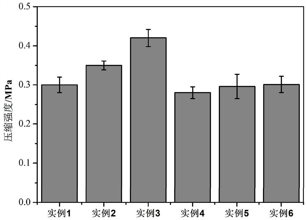
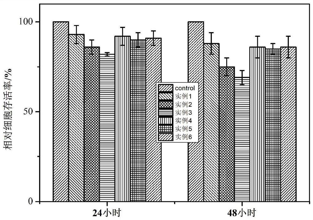
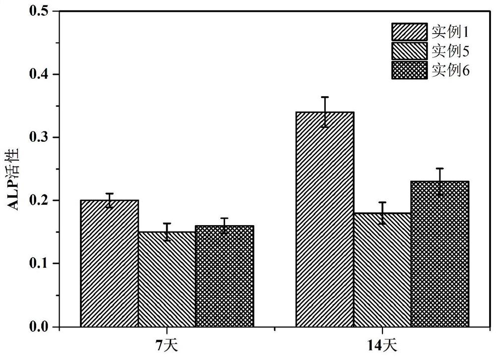
[0052] (1) Establishment of osteochondral scaffold model: According to the principles of bionics and the size and structure of bone defects, use 3D modeling software to design a bionic bone scaffold model, and finally export the model to a 3D printer in STL format. The model in this example is: diameter 10mm, height 7mm The cylindrical gradient scaffolds are composed of cartilage layer, calcified cartilage layer and subchondral bone layer from top to bottom. The thickness of the lower bone layer is 3.5 mm, the pore diameter is 200-500 μm, and the pores are interconnected.
[0053] (2) Synthetic preparation of GelMA: Weigh 5g of gelatin with an analytical balance, dissolve it in 50mL of PBS solution (pH 7.4, 0.01M), and dissolve it completely at 60°C to obtain 50mL of gelatin solution, place the gelatin solution on Stir on a constant temper...
Embodiment 2
[0060] A method for preparing a gradient osteochondral biomimetic scaffold integrated with diagnosis and treatment, which is basically the same as in Example 1, except that the sol ratio of each layer is different:
[0061] Cartilage lamellar sol: Weigh quercetin and GelMA, dissolve and mix them evenly with LAP solution, the final concentrations of the two are 0.03% (w / w) and 18% (w / w) respectively to obtain cartilage lamellar sol, namely Example 2 cartilage lamellar sol.
[0062] Cartilage calcification layer sol: Weigh quercetin, GelMA and Fe-HAP, dissolve and mix them evenly with LAP solution, and the final concentrations of the three are 0.03% (w / w), 23% (w / w) and 3% (w / w) respectively. / w), namely get example 2 cartilage calcification layer sol.
[0063] Subchondral bone layer sol: weigh quercetin, GelMA and Fe-HAP, dissolve and mix them evenly with LAP solution, the final concentrations of the three are 0.03% (w / w), 28% (w / w) and 3% respectively (w / w), promptly obtains...
Embodiment 3
[0065] A method for preparing a gradient osteochondral biomimetic scaffold integrated with diagnosis and treatment, which is basically the same as in Example 1, except that the sol ratio of each layer is different:
[0066] Cartilage lamellar sol: Weigh quercetin and GelMA, dissolve and mix them evenly with LAP solution, the final concentrations of the two are 0.03% (w / w) and 20% (w / w) respectively to obtain cartilage lamellar sol, namely Example 3 cartilage lamellar sol.
[0067] Cartilage calcification layer sol: Weigh quercetin, GelMA and Fe-HAP, dissolve and mix them evenly with LAP solution, and the final concentrations of the three are 0.03% (w / w), 25% (w / w) and 3% (w / w) respectively. / w), namely get example 3 cartilage calcification layer sol.
[0068] Subchondral bone layer sol: Weigh quercetin, GelMA and Fe-HAP, use LAP solution to dissolve and mix evenly, and the final concentrations of the three are 0.03% (w / w), 30% (w / w) and 3% ( w / w), promptly get example 3 subc...
PUM
| Property | Measurement | Unit |
|---|---|---|
| thickness | aaaaa | aaaaa |
| thickness | aaaaa | aaaaa |
| thickness | aaaaa | aaaaa |
Abstract
Description
Claims
Application Information
 - R&D
- Intellectual Property
- Life Sciences
- Materials
- Tech Scout
- Unparalleled Data Quality
- Higher Quality Content
- 60% Fewer Hallucinations
Browse by: Latest US Patents, China's latest patents, Technical Efficacy Thesaurus, Application Domain, Technology Topic, Popular Technical Reports.
© 2025 PatSnap. All rights reserved.Legal|Privacy policy|Modern Slavery Act Transparency Statement|Sitemap|About US| Contact US: help@patsnap.com


