Shigella flexneri microphage SGF3 and application thereof
A technology of Shigella flexneri and Shigella, applied in the direction of phage, virus/phage, application, etc., can solve the problems of poor antibacterial effect, improve the breadth and effectiveness, and expand the host spectrum Effect
- Summary
- Abstract
- Description
- Claims
- Application Information
AI Technical Summary
Problems solved by technology
Method used
Image
Examples
Embodiment 1
[0020] The separation and purification of embodiment 1 phage
[0021] The sewage sample used in the experiment of the present invention was collected in the west area of Yanqi Lake Campus of the University of Chinese Academy of Sciences in Beijing in November 2019, and used as a water sample for the isolation of bacteriophage.
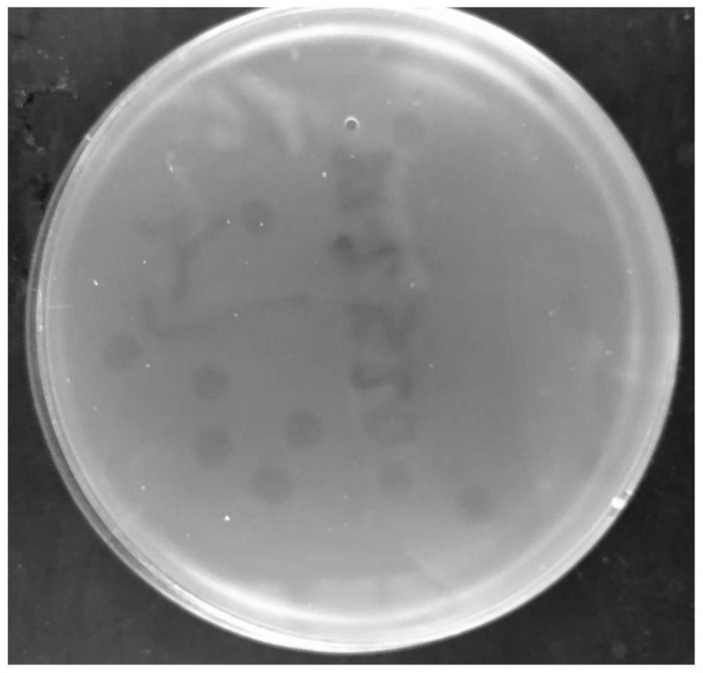
[0022] Centrifuge the sewage sample for 15min, take 25mL of sewage sterilized by filtration with a 0.22μm microporous membrane, add CaCl 2 Mother liquor to a final concentration of 1mmol / L, let it stand still, add 20ml NB liquid medium, then add 2.0ml of logarithmic phase host bacterial suspension, mix well in a 50ml centrifuge tube, let stand for 15min, shake overnight at 37°C ( 10h). The next day, add 2 mL of chloroform, centrifuge at 8,000 g for 30 min at 4°C, and the supernatant is the stock solution containing the phage of the host bacterium. Use the spot-test method to identify whether the stock solution contains phages. Take an appropriate a...
Embodiment 2
[0024] Morphological observation of embodiment 2 bacteriophage
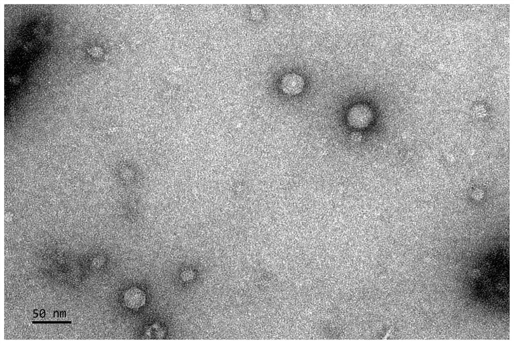
[0025] Take 20 μL of the phage suspension and drop it on the copper grid, wait for its natural precipitation for 10 minutes, blot it dry from the side with dry filter paper, let it air for about 1 minute, add 1 drop of 1% uranyl acetate on the copper grid, stain for 2 minutes, and then use it carefully to dry Blot the excess dye from the side of the filter paper, let it dry naturally in the dark for 30 minutes, and observe it with a transmission electron microscope (JEM-1400).
[0026] The results of transmission electron microscopy are shown in figure 2 , the results showed that the phage SGF3 has a polyhedral structure with a diameter of 28±4nm. According to the eighth report of the International Organization for Taxonomy of Viruses (ICTV) virus classification, the phage SGF3 can be preliminarily classified as a microphage family.
Embodiment 3
[0027] Example 3 Determination of Phage SGF3 Host Spectrum
[0028] The 14 strains in our laboratory were used to determine the host spectrum of SGF3, including Shigella, Escherichia coli, Staphylococcus aureus and Salmonella. The infection of SGF3 to each strain was detected by the double-layer plate method, and each experiment was repeated 3 times, with two parallel samples each time. The infection status of phages to the strains is shown in Table 1.
[0029] Table 1
[0030]
[0031] Features: After the phage infects the bacterial strain, (+) has phage plaques, (-) has no phage plaques.
[0032] CDC is the Chinese Center for Disease Control and Prevention; CGMCC is the Chinese General Microorganism Culture Collection and Management Center.
[0033] It can be seen from Table 1 that SGF3 only specifically lysed the host strain 1.10599, and had no lytic effect on other strains.
PUM
| Property | Measurement | Unit |
|---|---|---|
| Incubation period | aaaaa | aaaaa |
Abstract
Description
Claims
Application Information
 - R&D
- Intellectual Property
- Life Sciences
- Materials
- Tech Scout
- Unparalleled Data Quality
- Higher Quality Content
- 60% Fewer Hallucinations
Browse by: Latest US Patents, China's latest patents, Technical Efficacy Thesaurus, Application Domain, Technology Topic, Popular Technical Reports.
© 2025 PatSnap. All rights reserved.Legal|Privacy policy|Modern Slavery Act Transparency Statement|Sitemap|About US| Contact US: help@patsnap.com


