Method for identifying avian leukosis virus and chicken infectious anemia virus through visual double LAMP

A technology for avian leukosis virus and chicken infectious anemia, which is applied in biochemical equipment and methods, recombinant DNA technology, and microbial determination/inspection, etc., and can solve the problems of increased amplicon contamination risk, deviation of Tm value, laboratory contamination, etc. problem, to achieve the effect of good specificity, low pollution and high sensitivity

Active Publication Date: 2021-06-25
GUANGXI VETERINARY RES INST
View PDF4 Cites 2 Cited by
  • Summary
  • Abstract
  • Description
  • Claims
  • Application Information

AI Technical Summary

Problems solved by technology

At present, there are three domestic research reports on the application of double LAMP that can realize the real differential diagnosis. One is the specific sequence recognition method based on the enzyme digestion of the amplified product. Enzyme site, after the constant temperature amplification, the amplified product is digested, and then the different products are distinguished according to the different positions of the digested product displayed on the agarose gel, but this method takes a long time. And it needs to open the cover to handle the product, which increases the risk of amplicon contamination
The second is to rely on a fluorescent PCR instrument to determine what kind of pathogen it is based on the different Tm values ​​of the characteristic peaks in the melting curve of the amplification product. This method may have different Tm values ​​for the amplification products of templates from different sources of the same pathogen. The Tm value that leads to the result is biased, so this method has certain limitations
The third is to introduce fluorescent groups with different emission wavelengths at the 5' end of the internal primer FIP. After electrophoresis of the amplified products, the amplified products of different pathogens emit different lights under different emission wavelength channels to distinguish Different pathogens, but this method also needs to open the cover for electrophoresis, which is easy to cause laboratory pollution

Method used


Image

  • Method for identifying avian leukosis virus and chicken infectious anemia virus through visual double LAMP
  • Method for identifying avian leukosis virus and chicken infectious anemia virus through visual double LAMP
  • Method for identifying avian leukosis virus and chicken infectious anemia virus through visual double LAMP

Examples


Embodiment 1

[0095] Embodiment 1, the design of double LAMP primer and the preparation of positive plasmid

[0096] 1. Design of primers and probes

[0097] According to the conserved sequence of the pol gene and CIAV VP2 gene of ALV (subtypes A, B, and J) in GenBank, LAMP primers were designed using MEGA 4.0 and Primer Explorer V4 online primer design software, and the outer primers included ALV-F3, ALV-B3 , CAV-F3 and CAV-B3, internal primers include ALV-FIP, ALV-BIP, CIAV-FIP and CIAV-BIP, internal primers FIP=F1c+F2, BIP=B1c+B2; Utilize PrimerPrimer5.0 in ALV and Two probes were designed between F1c and B1c of CIAV, respectively ALV-probe, 5' end labeled with FAM fluorophore, 3' end labeled with BHQ3 quencher group; CIAV-Probe, 5' end labeled with CY 5 fluorophore group, the 3' end is labeled with the BHQ3 quencher group. The sequences of primers and probes are shown in Table 1. The sequences of inner primers and outer primers were synthesized by Huada Gene Biotechnology (Shenzhen) C...

Embodiment 2

[0110] Embodiment 2, establishment of double LAMP method

[0111] 1. Reaction system

[0112] 2 μL DNA / cDNA template, 10 μL 2×Reaction Mix, 0.8 μL Bst DNApolymerase, 0.8 μL each of internal primers ALV-FIP, ALV-BIP, CIAV-FIP and CIAV-BIP (both working concentrations are 40 μmol / L), external primer ALV -0.4 μL each of F3, ALV-B3, CIAV-F3 and CIAV-B3 (working concentration is 5 μmol / L), 0.4 μL of ALV-Probe (working concentration is 0.5 μmol / L), CIAV-Probe (working concentration is 0.5 μmol / L) 0.8μL plus ddH 2 After making up 20 μL of O, the reaction tube was placed in a thermostat or a Loopamp LA-320C real-time turbidimeter to react at 62°C for 60 minutes, and inactivated at 80°C for 5 minutes. After the test, the reaction tube was directly placed in a multicolor fluorescence imaging system for determination.

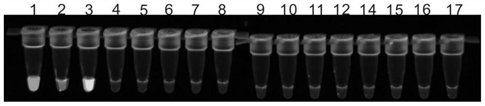
[0113] 2. Visual double LAMP result judgment

[0114] Visual double LAMP After judging the amplification result by monitoring the turbidity with a real-time turbidimet...

Embodiment 3

[0115] Embodiment 3, specificity test

[0116] Samples to be tested: positive ALV (mixed template of A subgroup, B subgroup and J subgroup), CIAV, ALV and CIAV mixed template, FAdV-4, ARV, NDV, AIV-H5, AIV-H7, AIV-H9 , AEV, REV, IBV, IBDV and MDV cDNA or DNA templates.

[0117] Using the sample to be tested as a template, perform double LAMP amplification according to the method established in Example 2.

[0118] Set RNase-free water as a negative control.

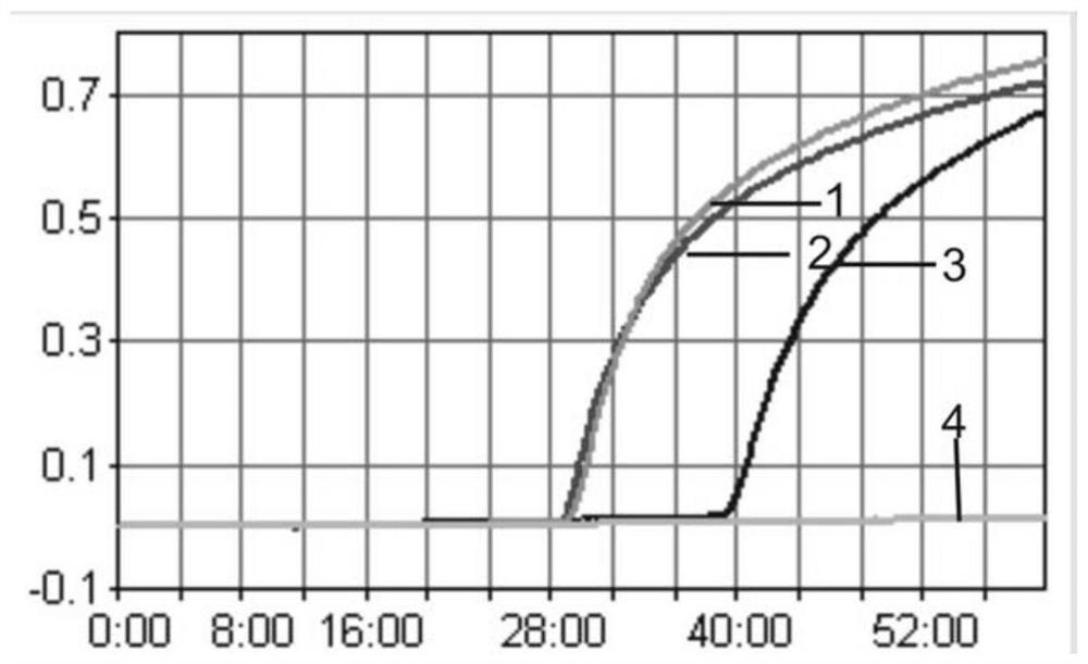
[0119] The cDNA of positive ALV and the DNA of CIAV are used as templates, RNase-free water is set as a negative control, and the reaction tube is placed in a Loopamp LA-320C real-time turbidimeter to react, and the results show that the cDNA of ALV and the DNA of CIAV are amplified ( figure 1 ). The positive ALV (mixed template of A subgroup, B subgroup and J subgroup), CIAV, ALV and CIAV mixed template, FAdV-4, ARV, NDV, AIV-H5, AIV-H7, AIV-H9, AEV, cDNA or DNA of REV, IBV, IBDV and MDV were used as templates, and RN...


PUM


Abstract

The invention discloses a method for identifying avian leukosis virus and chicken infectious anemia virus through visual double LAMP. The invention provides a single-stranded DNA group for identifying avian leukosis virus and chicken infectious anemia virus through visual double LAMP. The single-stranded DNA group consists of single-stranded DNA molecules as shown in SEQ ID No.1-10. According to the invention, the visual double LAMP method for identifying and detecting the ALV and the CIAV is successfully established. The double LAMP can identify and diagnose the ALV and the CIAV in the same reaction tube, has the advantages of good specificity, high sensitivity, small pollution, convenience, rapidness and the like, a detection result can be directly observed by naked eyes, the result is judged according to the color of a reaction product, and the double LAMP is suitable for clinical rapid screening of the ALV and the CIAV.

Description

technical field [0001] The invention relates to the field of poultry virus detection, in particular to a method for distinguishing avian leukosis virus and chicken infectious anemia virus by visual double LAMP. Background technique [0002] At present, the phenomenon of double and multiple infection of immunosuppressive virus in chicken flocks in my country is common. Avian leukemia (Avian leukemia, AL) and Chicken Infectious Anemia (Chicken Infectious Anemia, CIA) are two immunosuppressive diseases of chickens. When CIAV and ALV are infected alone or co-infected, they can significantly reduce the immune effect of the vaccine, which may The chickens cannot effectively resist the infection of the virus and develop other diseases, which in turn affects the health and economic benefits of the breeding industry. Studies by Wang Xian and others have shown that CIAV infection is common in domestic chicken flocks, and CIAV is often co-infected with ALV, Marek's disease virus (MDV)...

Claims


Application Information

Patent Timeline
IPC IPC(8): C12N15/11C12Q1/70C12Q1/6844
CPCC12Q1/701C12Q1/6844C12Q2600/16C12Q2600/166C12Q2531/119C12Q2537/143C12Q2563/107C12Q2545/113Y02A50/30
Inventor 谢芝勋张民秀谢志勤谢丽基张艳芳邓显文曾婷婷罗思思
Owner GUANGXI VETERINARY RES INST
Why Eureka
Try Eureka