
Arginine fluorescent probe and preparation method and application thereof

An arginine, optical probe technology, applied in the field of optical probes, which can solve the problems of in situ, real-time, dynamic, high-throughput and high spatiotemporal resolution detection of non-viable cells and subcellular organelles

Pending Publication Date: 2021-09-03
EAST CHINA UNIV OF SCI & TECH
View PDF5 Cites 1 Cited by
  • Summary
  • Abstract
  • Description
  • Claims
  • Application Information

AI Technical Summary

Problems solved by technology

[0006] However, there are great defects in these detection methods in the study of living cells, which require time-consuming sample processing processes: cell disruption, separation, extraction and purification, etc., and cannot be used in situ, real-time, dynamic, high-pass in living cells and subcellular organelles. volume and high spatiotemporal resolution

Method used


Image

  • Arginine fluorescent probe and preparation method and application thereof
  • Arginine fluorescent probe and preparation method and application thereof
  • Arginine fluorescent probe and preparation method and application thereof

Examples


Embodiment 1

[0136] Example 1: Arginine-binding protein particles

[0137] The STM4351 gene in the Agrobacterium agrobacterium gene was amplified by PCR, and the PCR product was recovered after gel electrophoresis and digested with BamHI and EcoRI, and the pET28a vector was subjected to the same double digestion. After ligation with T4 DNA ligase, the ligation product was transformed into Trans5a, and the transformed Trans5a was spread on an LB plate (kanamycin 100ug / mL), and cultured at 37°C overnight. After plasmid extraction of the growing Trans5a transformants, PCR identification was performed. After the positive plasmid is correctly sequenced, the subsequent plasmid construction is carried out.

Embodiment 2

[0138] Example 2: Expression and detection of cpYFP optical probes at different insertion sites

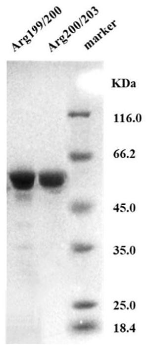
[0139] In this example, based on pET28a-STM4351, the following sites were selected to insert cpYFP according to the crystal structure of arginine-binding protein, and the corresponding plasmids were obtained: 103 / 104, 103 / 105, 103 / 106, 103 / 107, 103 / 108, 103 / 109, 103 / 110, 103 / 111, 104 / 105, 104 / 106, 104 / 107, 104 / 108, 104 / 109, 104 / 110, 104 / 111, 105 / 106, 105 / 107, 105 / 108, 105 / 109, 105 / 110, 105 / 111, 106 / 107, 106 / 108, 106 / 109, 106 / 110, 106 / 111, 107 / 108, 107 / 109, 107 / 110, 107 / 111, 108 / 109, 108 / 110, 108 / 111, 109 / 110, 109 / 111, 110 / 111, 197 / 198, 197 / 199, 197 / 200, 197 / 201, 197 / 202, 197 / 203, 197 / 204, 197 / 205, 197 / 206, 197 / 207, 197 / 208, 197 / 209, 198 / 199, 198 / 200, 198 / 201, 198 / 202, 198 / 203, 198 / 204, 198 / 205, 198 / 206, 198 / 207, 198 / 208, 198 / 209, 199 / 200, 199 / 201, 199 / 202, 199 / 203, 199 / 204, 199 / 205, 199 / 206, 199 / 207, 199 / 208, 199 / 209, 200 / 201, 200 / 202, 200 / 203, 200 / 204, 200 / 205, 200 / 206, 200...

Embodiment 3

[0145] Example 3, Expression and detection of cpGFP optical probes at different insertion sites

[0146] According to the method in Example 1, cpYFP was replaced by green fluorescent protein cpGFP, which was fused into an arginine-binding protein to construct an arginine green fluorescent protein fluorescent probe, which was expressed and detected according to the method in Example 2. The result is as image 3 As shown, the results of fluorescence detection showed that 104 / 110, 105 / 106, 105 / 109, 105 / 110, 105 / 111, 106 / 108, 106 / 109, 107 / 109, 107 had more than 2 times response to arginine / 111, 197 / 203, 198 / 201, 199 / 200, 199 / 202, 199 / 203, 199 / 204, 200 / 202, 200 / 203, 200 / 204, 200 / 205, 200 / 206, 201 / 202 , 201 / 205, 204 / 205 sites.


PUM


Abstract

The invention provides an arginine fluorescent probe. The arginine fluorescent probe comprises polypeptide B responding to arginine and fluorescent protein A expressing the arginine; the fluorescent protein A is inserted into the polypeptide B, the polypeptide B is divided into two upper and lower structural parts of B1 and B2, and a B1-A-B2 type probe structure is formed; similarly, truncated and site-specific mutagenesis optimized mutants are located at different positions, and the polypeptide B and the arginine are specifically bound to cause change of a fluorescent signal of the fluorescent protein A; and the polypeptide B is arginine binding protein and a mutant thereof. The arginine fluorescent probe is relatively small in protein molecular weight, easy to express, large in fluorescent dynamic change and good in specificity, can be expressed in different subcellular organelles of cells through gene operation, and can quantitatively detect the arginine in and out of the cells in a high-throughput mode.

Description

technical field [0001] The invention relates to the technical field of optical probes, in particular to an arginine optical probe and its preparation method and application. Background technique [0002] As one of the 20 kinds of natural amino acids, arginine was originally isolated and extracted from the plant lupine seedlings by Schlus in 1886. Its molecular structure has been clarified and can be artificially synthesized in the early 20th century. Arginine exerts biological functions in the form of physiologically active L-arginine in organisms. Arginine is not only a component of protein in the body, but also a precursor for the synthesis of various biologically active substances, such as polyamines and NO, etc., by stimulating the secretion of some hormones, it participates in biological processes such as endocrine regulation and body-specific immune regulation; In addition, it is also used as an intermediate in the urea cycle to relieve ammonia poisoning through the u...

Claims


Application Information

Patent Timeline
Patent Type & Authority Applications(China)
IPC IPC(8): C07K19/00C07K14/255C12N15/31G01N33/68
CPCC07K14/255G01N33/6806C07K2319/00C07K2319/60G01N33/6812C07K2319/21C07K14/001G01N33/533
Inventor 杨弋赵玉政李睿邹叶君黄立李写陈念
Owner EAST CHINA UNIV OF SCI & TECH
Learn More