

Staining method for living cell imaging
A technology of living cells and staining methods, which is applied in the field of cell staining and double staining using fluorescent biomarkers, which can solve the problems of inability to observe the shape of the nucleus, the inability to observe the cell shape, and the long staining process.
- Summary
- Abstract
- Description
- Claims
- Application Information
AI Technical Summary
Problems solved by technology
Method used
Image

Examples
Embodiment 1
[0081] Example 1: Tissue staining by sodium fluorescein and methylene blue double staining
[0082] 1.1 Mouse kidney staining
[0083] Using mice for tissue staining, the staining process is as follows:
[0084] 1. Mice were anesthetized by intraperitoneal injection of 1% sodium pentobarbital, and the anesthesia dose was 8–9 mL / g.
[0085] 2. Remove the body hair on the back of the mouse, cut the epidermis to expose the kidney, remove the kidney completely, fix the surface of the kidney with a blade, and remove the capsule on the surface of the kidney with scissors and tweezers under a microscope.
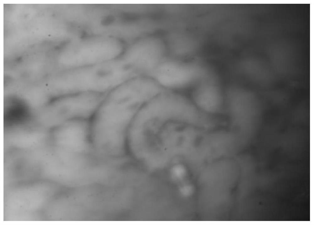
[0086] 3. Use a cotton swab to stop bleeding, apply 0.25% sodium fluorescein to the surface of the kidney for 2 minutes, wash it with normal saline three times; then apply 1% melanin staining solution for 2 minutes, wash it with normal saline three times after the time is up, Then place it under a microscope for observation at a wavelength of 470nm.
[0087] The results in Figur...
Embodiment 2
[0095] Example 2: Effects of double staining with different concentrations of fluorescein sodium and methylene blue staining
[0096] Use porcine kidney for tissue staining, the staining process is as follows:
[0097] 1. Purchase some fresh pig kidneys and refrigerate them at 4°C for later use.
[0098] 2. Clean the fresh pig kidney with clean water, and remove the membrane on the surface of the kidney with scissors and tweezers under a microscope.
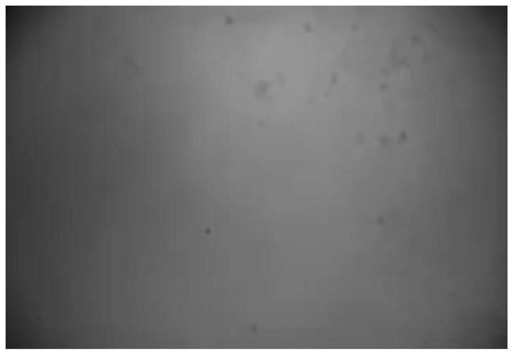
[0099] 3. Apply and stain the surface of the kidney with 0.1%, 0.25%, 0.5%, and 1% fluorescein sodium for one minute, then wash with normal saline three times; then stain with 0.5%, 1%, 2%, and 3% methylene blue respectively Apply and dye the surface of the liquid for one minute. After the time is up, wash it with physiological saline three times, and then observe it under a microscope with a wavelength of 470nm.
[0100] Figure 3 is a comparison of the imaging effects of pig kidneys stained with different concentrations of sod...
Embodiment 3
[0102] Example 3: 5-ALA and Acridine Yellow Double Staining Method for Tissue Staining
[0103] Pig liver was used for tissue staining, and the staining process was as follows:
[0104] 1. Purchase some fresh pig livers and refrigerate them at 4°C for later use.
[0105] 2. Clean the fresh pig liver with clean water, and remove the surface film of the liver with scissors and tweezers under a microscope.
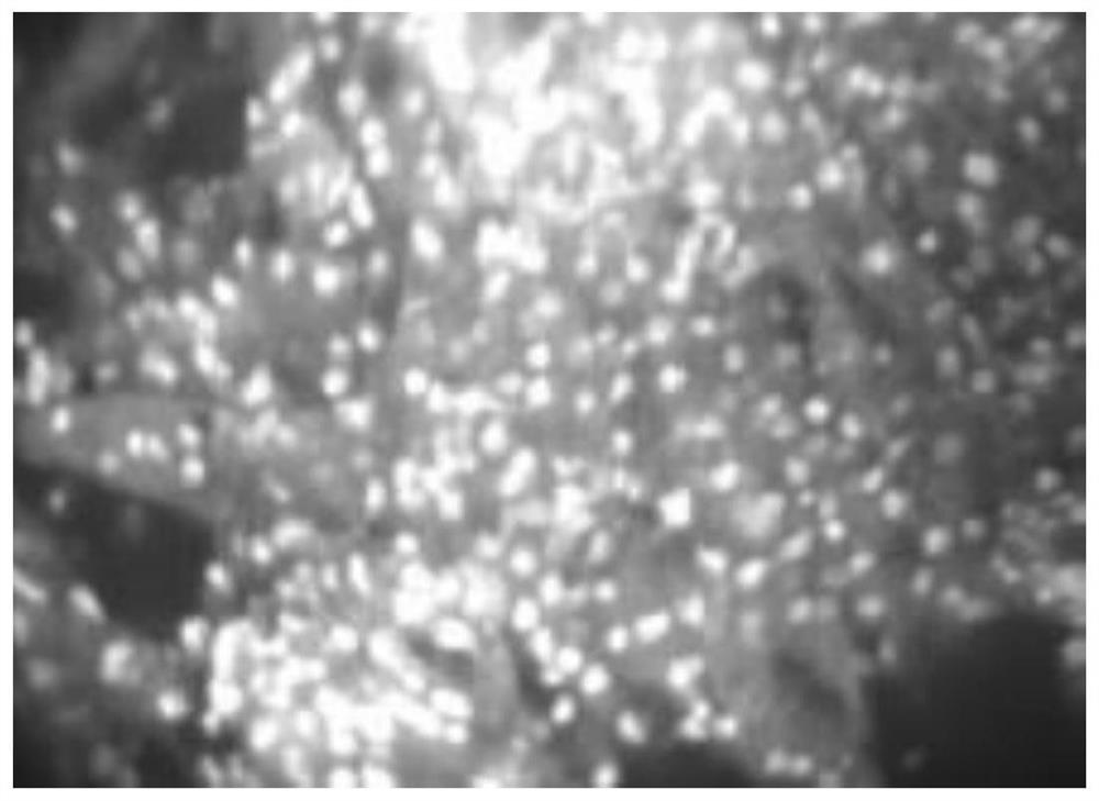
[0106] 3. Dye the surface of the liver with 0.05% 5-ALA for 3 minutes, wash it with normal saline three times; then apply and dye the surface with 1% acridine yellow staining solution for 2 minutes, wash it with normal saline three times after the time is up, and then put it in Observed under a microscope at a wavelength of 635nm.
[0107] Figure 4 The imaging results of double-staining with 5-ALA and acridine yellow for pig liver can clearly see the outline of the stained liver cells and the shape of the nucleus.
PUM
| Property | Measurement | Unit |
|---|---|---|
| Excitation wavelength | aaaaa | aaaaa |
Abstract
Description
Claims
Application Information