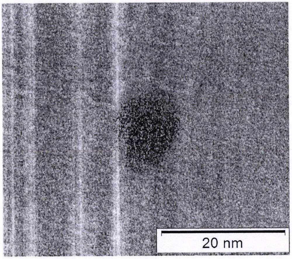
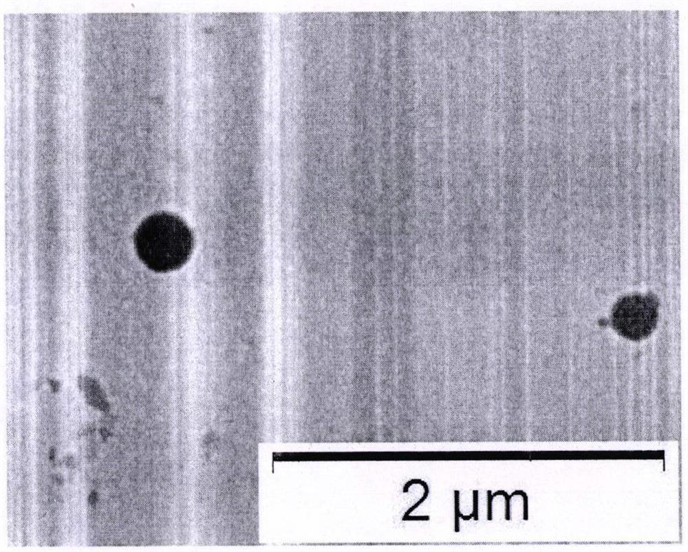
Nano gene drug transporter
A gene drug and carrier technology, applied in the field of spherical calcium phosphate nano-gene drug delivery carrier, can solve the problems of low shape and external load
- Summary
- Abstract
- Description
- Claims
- Application Information
AI Technical Summary
Problems solved by technology
Method used
Image
Examples
example 1
[0024] 1) Preparation of green fluorescent carbon quantum dots: Cysteine and sucrose in equimolar ratio were added to a round bottom flask, and 20 mL of 0.1 mol / L sodium hydroxide solution was added. Stir and reflux at 80° C. for 24 hours to obtain an orange-yellow liquid. After cooling to room temperature, centrifuge to remove larger particles (11,000 rpm, 15 minutes), and collect the supernatant solution for later use. The particle size of carbon quantum dots is 1-9nm.
[0025] 2) Prepare a phosphate buffer solution with a pH value of 11; prepare a calcium chloride solution according to a calcium-phosphorus ratio of 1.7. Prepare 2mmol / L cetyltrimethylammonium bromide solution.
[0026] 3) Preparation of spherical calcium phosphate nanoparticles: add 2 mL of the above-prepared carbon quantum dot solution, 2 mL of cetyltrimethylammonium bromide solution, and 15 mL of distilled water to prepare a mixed solution in a round bottom flask, heat and stir in a water bath at 80°C ...
example 2
[0028] 1) Preparation of nucleic acid crystal nucleus: use protamine sulfate solution to polycondense nucleic acid into a spherical shape;
[0029] 2) Prepare a phosphate buffer solution with a pH value of 9; prepare a calcium nitrate solution according to a calcium-phosphorus ratio of 1.7. Prepare 2mmol / L tetradecyltrimethylammonium bromide solution.
[0030] 3) Preparation of spherical calcium phosphate nanoparticles: Add 0.5mL of the above-prepared nucleic acid solution, 0.5mL of tetradecyltrimethylammonium bromide solution, and 4mL of distilled water to prepare a mixed solution in a sterile round-bottomed flask, and place in a water bath at 40°C Heat and stir for 20min. Afterwards, 2 mL of phosphate buffer solution was added to the reaction system, and after heating and stirring for 20 min, 2 mL of calcium nitrate solution was added to the system, followed by condensation and reflux for 24 h. After the synthesis reaction is completed, the resulting suspension is washed w...
example 3
[0032] 1) Preparation of cisplatin-polylactic acid crystal nucleus: polylactic acid is used to wrap cisplatin in the way of water-in-oil-in-water, and the pellets of about 20 nm are made.
[0033] 2) Prepare a phosphate buffer solution with a pH value of 10; prepare a calcium nitrate solution according to a calcium-phosphorus ratio of 1.7. Prepare 2mmol / L octadecyltrimethylammonium bromide solution.
[0034] 3) Preparation of spherical calcium phosphate nanoparticles: suspend the above-mentioned cisplatin-polylactic acid nanospheres in 0.5 mL aqueous solution, add 0.5 mL of octadecyltrimethylammonium bromide solution, and 4 mL of distilled water to prepare a mixed solution in the absence of In a round-bottomed flask, heat and stir in a water bath at 50°C for 20 min. Afterwards, 2 mL of phosphate buffer solution was added to the reaction system, and after heating and stirring for 20 min, 2 mL of calcium nitrate solution was added to the system, followed by condensation and ref...
PUM
| Property | Measurement | Unit |
|---|---|---|
| size | aaaaa | aaaaa |
| size | aaaaa | aaaaa |
| size | aaaaa | aaaaa |
Abstract
Description
Claims
Application Information
 - R&D
- Intellectual Property
- Life Sciences
- Materials
- Tech Scout
- Unparalleled Data Quality
- Higher Quality Content
- 60% Fewer Hallucinations
Browse by: Latest US Patents, China's latest patents, Technical Efficacy Thesaurus, Application Domain, Technology Topic, Popular Technical Reports.
© 2025 PatSnap. All rights reserved.Legal|Privacy policy|Modern Slavery Act Transparency Statement|Sitemap|About US| Contact US: help@patsnap.com


