3D printing porous surface zirconium oxide dental implant and design and processing method thereof

A 3D printing, porous surface technology, applied in the field of medical devices, can solve the problems of poor surface treatment effect and osseointegration performance, and achieve the effect of facilitating cell migration, promoting osseointegration, and good biocompatibility

Pending Publication Date: 2022-02-08
PEKING UNIV SCHOOL OF STOMATOLOGY
View PDF0 Cites 1 Cited by
  • Summary
  • Abstract
  • Description
  • Claims
  • Application Information

AI Technical Summary

Problems solved by technology

[0008] The basic idea of ​​the present invention is: according to different actual clinical needs, a series of porous zirconia dental implants are produced by using 3D printing technology, the surface layer has high porosity, the core has high density, and the porosity and pore size of the porous structure can be adjusted , can provide a method for the optimal design of porous implants, and is expected to solve the clinical problems of conventional zirconia implant surface treatment effects and poor osseointegration performance

Method used


Image

  • 3D printing porous surface zirconium oxide dental implant and design and processing method thereof
  • 3D printing porous surface zirconium oxide dental implant and design and processing method thereof
  • 3D printing porous surface zirconium oxide dental implant and design and processing method thereof

Examples


Embodiment 1

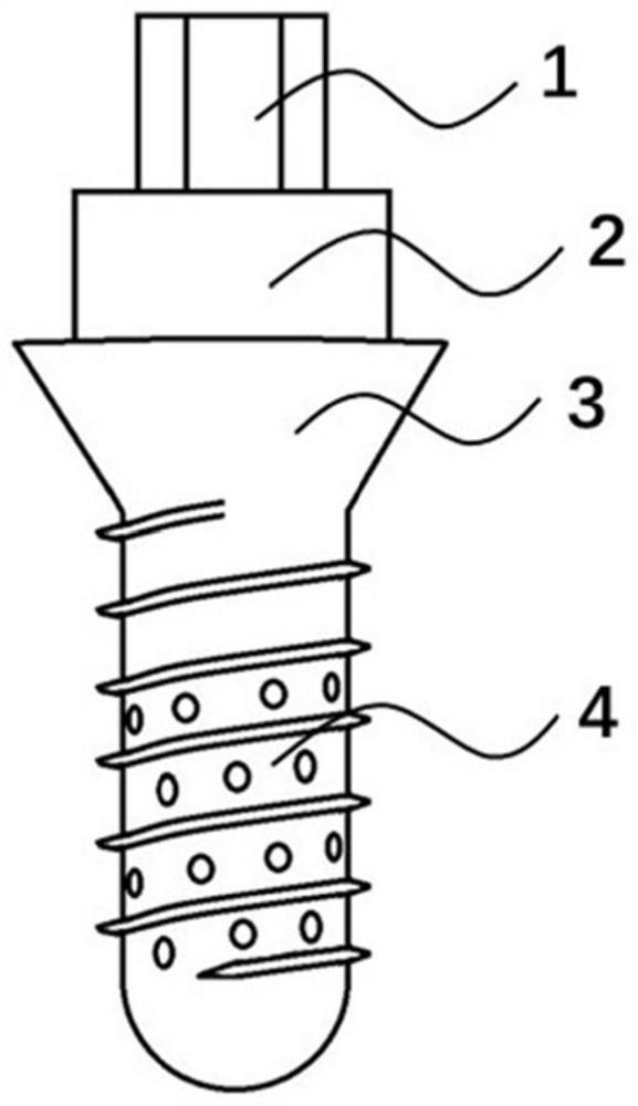
[0108] Porous surface design: the diameter of the implant is 4.5 mm, the porous structure is evenly distributed from 3 mm below the nominal bone plane to the end thread, the pore diameter is 100 μm, the hole depth is 150 μm, and 4 rows of porous bands can be arranged between adjacent threads. The perforated belts are arranged parallel to the rotation of the threads.

[0109] Material: The main components of the printing material are 3Y-TZP (polycrystalline tetragonal zirconia stabilized with 3% mol yttria) and photosensitive resin.

[0110] Printing parameters: printing angle 0°, printing layer thickness 25 μm, base support height of support structure 1.0 mm, gap with implant 0.2 mm, draft angle 10°.

[0111] Cleaning, drying, and degreasing and sintering: Remove the supporting structure from the printed implant, ultrasonically wash it in isopropanol solution for 10-15s, repeat 2-3 times, vacuum dry for 1min, and degrease and sinter for 52 hours.

[0112] Test data: static st...

Embodiment 2

[0115] Porous surface design: the diameter of the implant is 4.5 mm, the porous structure is evenly distributed from 3 mm below the nominal bone plane to the end thread, the pore diameter is 400 μm, the hole depth is 600 μm, and a row of porous bands can be arranged between adjacent threads. The perforated belts are arranged parallel to the direction of thread rotation.

[0116] Material: The main components of the printing material are 3Y-TZP (polycrystalline tetragonal zirconia stabilized with 3% mol yttria) and photosensitive resin.

[0117] Printing parameters: printing angle 0°, printing layer thickness 25 μm, base support height of support structure 1.0 mm, gap with implant 0.2 mm, draft angle 10°.

[0118] Cleaning, drying, and degreasing and sintering: Remove the supporting structure from the printed implant, ultrasonically wash it in isopropanol solution for 10-15s, repeat 2-3 times, vacuum dry for 1min, and degrease and sinter for 52 hours.

[0119] Test data: stati...

Embodiment 3

[0122] Porous surface design: the diameter of the implant is 4.5 mm, the porous structure is evenly distributed from 6 mm below the nominal bone plane to the end thread, the pore diameter is 400 μm, and the hole depth is 600 μm. A row of porous bands can be arranged between adjacent threads. The perforated belts are arranged parallel to the rotation of the threads.

[0123] Material: The main components of the printing material are 3Y-TZP (polycrystalline tetragonal zirconia stabilized with 3% mol yttria) and photosensitive resin.

[0124] Printing parameters: printing angle 0°, printing layer thickness 25 μm, base support height of support structure 1.0 mm, gap with implant 0.2 mm, draft angle 10°.

[0125] Cleaning, drying, and degreasing and sintering: Remove the supporting structure from the printed implant, ultrasonically wash it in isopropanol solution for 10-15s, repeat 2-3 times, vacuum dry for 1min, and degrease and sinter for 52 hours.

[0126] Test data: static str...


PUM

PropertyMeasurementUnit
diameteraaaaaaaaaa
depthaaaaaaaaaa
heightaaaaaaaaaa

Abstract

The invention relates to a 3D printing porous surface zirconium oxide dental implant and a design and processing method thereof. The 3D printing porous surface zirconium oxide dental implant comprises an intraosseous porous implant and a supraosseous abutment, and the porous surface implant is produced and processed by 3D printing; the porous structure of the porous surface implant is adjustable and controllable in pore shape, diameter and porosity; the 3D printing adopts a three-dimensional light curing method or a digital light processing method. The method comprises the following steps: forming a dental implant structure with high surface porosity and high core density by using a computer-aided design technology, and processing by using a 3D printing technology to obtain the porous surface zirconium oxide implant with a controllable pore structure, so that the surface design and processing of the zirconium oxide implant can be optimized, and the bone bonding performance can be improved; the individuation degree of planting restoration treatment is improved.

Description

technical field [0001] The invention relates to the technical field of medical devices, in particular to a 3D printed porous surface zirconia dental implant. Background technique [0002] Oral implant restoration has become one of the best treatment methods for dentition defects. Zirconia is a biologically inert ceramic material with beautiful and natural color, good biocompatibility and chemical stability. Compared with pure titanium with the same surface roughness, zirconia is not easy to accumulate plaque in the mouth, which is beneficial to the health of the soft tissue around the implant. With the continuous optimization of the properties of zirconia materials and the gradual improvement of digital processing technology, zirconia has a good application prospect as a suitable choice of implant materials. [0003] At present, the zirconia implants used in the market are mainly processed by injection molding, and the degree of personalization is low. Implants made of a s...

Claims


Application Information

Patent Timeline
Patent Type & Authority Applications(China)
IPC IPC(8): A61C8/00A61C13/00A61C13/08C04B35/48C04B35/622C04B38/00B33Y10/00B33Y50/02
CPCA61C8/0037A61C13/08A61C13/0019C04B38/00C04B35/48C04B35/622B33Y10/00B33Y50/02
Inventor 张磊丁茜赵健霄马全诠孙玉春李文锦高远陈杰
Owner PEKING UNIV SCHOOL OF STOMATOLOGY
Why Eureka
Try Eureka