Novel crown RBD protein fluorescent microsphere composite preparation
A technology of composite preparation and fluorescent microspheres, applied in the field of saturated competition, can solve the problems of affecting the performance of detection reagents and the inability to perform detection, and achieve the effect of improving detection sensitivity and maintaining antigen activity.
- Summary
- Abstract
- Description
- Claims
- Application Information
AI Technical Summary
Problems solved by technology
Method used
Image
Examples
Embodiment 1
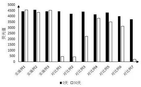
[0052] A new crown RBD protein fluorescent microsphere composite preparation, the preparation method is as follows:
[0053] (1) Weigh 10mg ovalbumin and add it to phosphate buffered saline (0.02-0.1M PBS, pH 7.2-7.4), then add 10-50mg / mL 1-ethyl-(3-dimethylaminopropyl ) Carbodiimide (EDC), avoid light reaction for 15-30min;
[0054] (2) Weigh 10 mg of bovine serum albumin and 10 mg of hemocyanin into the activated ovalbumin solution;
[0055] (3) Dialyze with phosphate buffered saline (0.02-0.1M PBS, pH 7.2-7.4) to remove EDC;
[0056] (4) After affinity purification, the product whose molecular weight was the sum of the molecular weights of the three proteins was collected to obtain coupling protein 1 (BSA-OVA-KLH);
[0057] (5) Weigh 2.5g trehalose, 0.9g NaCl, 0.05g PEG4000, 0.05g Tween 20, 0.05gproclin 300, 0.24g Tris, 100g process water, add coupling protein 1 to make the final concentration 1%, adjust the pH to 8.5, to obtain the compound preparation.
Embodiment 2
[0059] A new crown RBD protein fluorescent microsphere composite preparation, the preparation method is as follows:
[0060] (1) Weigh 10mg bovine serum albumin and add it to phosphate buffered saline (0.02-0.1M PBS, pH 7.2-7.4), then add 10-50mg / mL 1-ethyl-(3-dimethylaminopropyl base) carbodiimide (EDC), avoid light reaction for 15-30min;
[0061] (2) Weigh 10 mg of silk fibroin and add it to the activated bovine serum albumin solution;
[0062] (3) Dialyze with phosphate buffered saline (0.02-0.1M PBS, pH 7.2-7.4) to remove EDC;
[0063] (4) After affinity purification, the product whose molecular weight was the sum of the molecular weights of the two proteins was collected to obtain coupling protein 2 (BSA-SF).
[0064] (5) Weigh 2.5g trehalose, 0.9g NaCl, 0.05g PEG4000, 0.05g Tween 20, 0.05gproclin 300, 0.24g Tris, 100g process water, add coupling protein 2 to make the final concentration 1%, adjust the pH to 8.5, to obtain the compound preparation.
Embodiment 3
[0066] A new crown RBD protein fluorescent microsphere composite preparation, the preparation method is as follows:
[0067] (1) Weigh 10mg of polylysine and add it to phosphate buffer (0.02-0.1M PBS, pH 7.2-7.4), then add 10-50mg / mL 1-ethyl-(3-dimethylaminopropyl base) carbodiimide (EDC), avoid light reaction for 15-30min;
[0068] (2) Weigh 10 mg bovine serum albumin and add it to the activated polylysine solution;
[0069] (3) Dialyze with phosphate buffered saline (0.02-0.1M PBS, pH 7.2-7.4) to remove EDC;
[0070] (4) After affinity purification, the product with higher protein concentration was collected to obtain coupling protein 3 (ε-PL-BSA);
[0071] (5) Weigh 5g sucrose, 1.3g MgCl 2 , 0.05g PEG4000, 0.02g Tetronin1307, 0.02g KroVin300M, 0.24g Tris, 100g process water, add coupling protein 3 to make the final concentration 1%, and adjust the pH to 8.5 to obtain a compound preparation.
PUM
 Abstract
Description
Claims
Application Information
 - R&D
- Intellectual Property
- Life Sciences
- Materials
- Tech Scout
- Unparalleled Data Quality
- Higher Quality Content
- 60% Fewer Hallucinations
Browse by: Latest US Patents, China's latest patents, Technical Efficacy Thesaurus, Application Domain, Technology Topic, Popular Technical Reports.
© 2025 PatSnap. All rights reserved.Legal|Privacy policy|Modern Slavery Act Transparency Statement|Sitemap|About US| Contact US: help@patsnap.com


