Cell cross contamination detection method and application thereof

A technology of cross-contamination and detection method, applied in the field of cell cross-contamination detection, can solve the problems of contamination and insufficient analysis ability of complex mixed cell system, and achieve the effects of high sensitivity, low allele coverage and high specificity.

Pending Publication Date: 2022-07-08
CHINA JILIANG UNIV
View PDF0 Cites 0 Cited by
  • Summary
  • Abstract
  • Description
  • Claims
  • Application Information

AI Technical Summary

Problems solved by technology

[0006] In view of the above-mentioned defects in the prior art, the present invention creatively utilizes the advantages of high polymorphism and low mutation rate of short DNA fragments (<200bp), and establishes a cell crossover method based on microhaplotype (microhaplotype) genetic markers. Contamination detection method and application, overcome the problem of insufficient analysis ability of complex mixed cell system due to shadow band and amplification imbalance of traditional STR typing method

Method used


Image

  • Cell cross contamination detection method and application thereof
  • Cell cross contamination detection method and application thereof
  • Cell cross contamination detection method and application thereof

Examples


Embodiment 1

[0042] Example 1 Simulated detection of cell cross-contamination based on mixed cell samples

[0043] 1. Cell culture

[0044] The DMEM medium containing 10% fetal bovine serum was used, and the cells were cultured in an incubator containing 5% carbon dioxide at 37° C., and the cells could be used after they had grown into monolayers.

[0045] 2. Simulate cell cross-contamination:

[0046] After the adherent cells are digested with trypsin, a single cell suspension is prepared, the corresponding cell concentration is determined, and the required corresponding single cell suspension volume is calculated according to the specific number of cells. (1:9, 1:19, 1:49, 1:99) were mixed and used as test samples for simulated contamination of cells.

[0047] 3. DNA extraction from simulated contaminated cells:

[0048] DNA extraction kits were used to extract the DNA of simulated contaminated cells according to the instructions, and the concentration was measured with a micro-nuclei...

Embodiment 2

[0064] Example 2 Simulated detection of cell cross-contamination based on mixed cell DNA samples

[0065] 1. Cell culture:

[0066] The DMEM medium containing 10% fetal bovine serum was used, and the cells were cultured in an incubator containing 5% carbon dioxide at 37° C., and the cells could be used after they had grown into monolayers.

[0067] 2. Cell DNA extraction:

[0068] The adherent cells were digested with trypsin to prepare a single-cell suspension; then, according to the instructions of the DNA extraction kit, the DNA of each cell was extracted separately, and the concentration was determined with a micro-nucleic acid protein detector.

[0069] 3. Simulate cell cross-contamination:

[0070] Determine the dilution ratio according to the measured DNA concentration, and dilute the extracted DNA from each cell to a concentration of 10ng / μL. Then, the extracted DNAs from different cells were mixed in different ratios (1:9, 1:19, 1:49, 1:99) to simulate cell cross-c...

Embodiment 3

[0079] Example 3 Comparison between the method of the present invention and the STR typing method

[0080] 1. Cell culture:

[0081] The DMEM medium containing 10% fetal bovine serum was used, and the cells were cultured in an incubator containing 5% carbon dioxide at 37° C. and used after the cells grew into a monolayer.

[0082] 2. Cell DNA sample preparation:

[0083] The cell line DNA extracted in the same batch in Example 1 was used, and mixed according to the ratio of different cell numbers (1:9, 1:19, 1:49, 1:99), as a detection sample for simulated cross-contamination of cells, and the sample concentration was ≥ 50ng / μL.

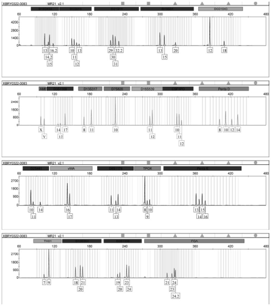
[0084] 3. STR analysis and identification:

[0085] Take an appropriate amount of simulated cross-contaminated cell DNA, use MicroreaderTM21 ID System to amplify 20 STR loci and sex identification loci, and use ABI 3730xl genetic analyzer to detect PCR products.

[0086] 4. STR information comparison:

[0087] The detection data information was pr...


PUM


Abstract

The invention discloses a cell cross contamination detection method which comprises the following steps: S1, preparing a cell cross contamination sample, and extracting cell DNA (Deoxyribose Nucleic Acid); s2, designing primers according to the site information to establish a multiple PCR amplification system; s3, performing high-throughput sequencing analysis after the library is successfully constructed; and S4, determining whether the cells are subjected to cross contamination or not according to a judgment standard. According to the invention, the micro-haplotype is analyzed through a high-throughput sequencing technology, all genotypes of SNP sites in a micro-haplotype composite system can be obtained, and the method has the advantages of high speed, high accuracy and reliability, integration and the like. The method is suitable for quality control of cell preparations and monitoring of quality of stored cell lines in a cell bank.

Description

technical field [0001] The invention relates to the technical field of biomedicine, in particular to a cell cross-contamination detection method and application. Background technique [0002] Cell cross-contamination refers to the contamination caused by the mixing of non-target cells from within or outside the species during the separation, culture and use of cells. In 1952, the first human-derived cell line HeLa cell line was successfully established, and cell culture has become an indispensable tool for the development of the biomedical industry since then. With the widespread use of cell culture, misidentification of cells or cross-contamination of cultures has gradually become a long-standing problem. According to statistics, there are more than 400 cell lines widely used in the world that have been confirmed to be misidentified or cross-contaminated, and at least 20% of the cell lines stored in laboratories in the United States, Europe and Asia are misidentified or cr...

Claims


Application Information

Patent Timeline
IPC IPC(8): C12Q1/6888C12Q1/6858C40B50/06C12N15/11
CPCC12Q1/6888C12Q1/6858C40B50/06C12Q2600/156C12Q2531/113C12Q2537/143C12Q2535/122C12Q2563/143C12Q2563/149Y02A50/30
Inventor 伍义行张彤彤朱羽婕廉润通叶子弘俞晓平马爱进赵同标
Owner CHINA JILIANG UNIV
Why Eureka
Try Eureka