Application of tirofiban in preparation of medicine for treating human respiratory syncytial virus infection

A technology of tirofiban and syncytial virus, which is applied in the field of anti-viral infection, can solve problems such as the lack of vaccines on the market, and achieve the effects of good general condition, normal feed intake, and active spirit

Active Publication Date: 2022-07-29
GUIZHOU MEDICAL UNIV
View PDF4 Cites 0 Cited by
  • Summary
  • Abstract
  • Description
  • Claims
  • Application Information

AI Technical Summary

Problems solved by technology

HRSV is the most important pathogen that causes viral pneumonia in children, and is also one of the important pathogens for lower respiratory tract infections in the...

Method used


Image

  • Application of tirofiban in preparation of medicine for treating human respiratory syncytial virus infection
  • Application of tirofiban in preparation of medicine for treating human respiratory syncytial virus infection
  • Application of tirofiban in preparation of medicine for treating human respiratory syncytial virus infection

Examples


Embodiment 1

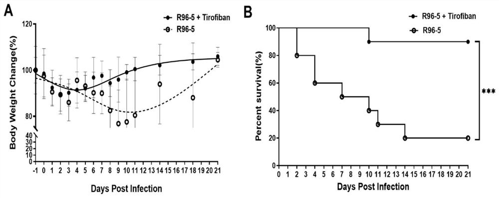
[0039] In vivo antiviral experiment: Mice were challenged 3 times with HRSV high-virulence strain R96-5 preserved in the Laboratory of Pathogen Biology, School of Basic Medicine, Guizhou Medical University, with a dose of 1 x 10 per challenge. 10 TCID 50 , at an interval of 12 hours, starting from the 1st day of the challenge, tirofiban was injected into the tail vein of mice at 50 μg / kg every day after the challenge to fight the virus infection, and injected continuously for 3 days; at the same time, a virus control group was set up. -5 mice were challenged 3 times with a dose of 1x 10 per challenge 10 TCID 50 , at an interval of 12 hours, starting from the first day of challenge, after daily challenge, the mice were injected with normal saline through the tail vein, and injected continuously for 3 days. The observation end point was 21 d after infection (the last challenge day was 0 d after infection).

[0040] During the observation period of 21 days after infection, we ...

Embodiment 2

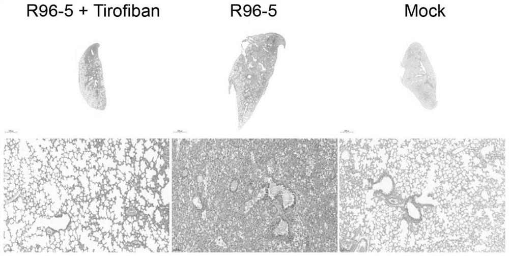
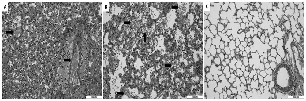
[0044] In the in vivo antiviral experiment of Example 1, mouse lungs were collected at different time points for pathological sections and H&E staining.

[0045] Mouse lung pathological sections, H&E staining results refer to figure 2 The results indicated that the local lung tissue of the tirofiban treatment group showed signs of mild to moderate viral pneumonia, and inflammatory cell infiltration was seen around small pulmonary blood vessels and bronchi, but most of the alveolar structures were intact, and no red blood cell leakage and thrombosis were found. . However, the HRSV infection group showed signs of severe viral pneumonia. A large number of inflammatory cells were infiltrated around small pulmonary blood vessels and bronchi, most of the alveolar structure was destroyed, and a large number of red blood cells leaked from small blood vessels to the alveolar space and pulmonary interstitium. Extensive thrombosis. figure 2 Middle Tirofiban: tirofiban; R96-5: HRSV hi...

Embodiment 3

[0050] In the in vivo antiviral experiment in Example 1, mouse lungs were collected at different time points for immunofluorescence staining experiments.

[0051] The expression level of HRSV F protein indicates the degree of virus infection, while the CD42b protein is a specific membrane protein of megakaryocytes and platelets. Figure 4 middle B and Figure 4 The results in middle G indicated that the degree of virus infection in the tirofiban treatment group was lower than that in the HRSV high strain R96-5 challenge group. Figure 4 Medium C and Figure 4 The results in middle H indicated that the number of virus platelets in the tirofiban treatment group was lower than that in the R96-5 challenge group, and the platelets of the mice in the HRSV high virus strain challenge group were distributed in alveolar tissue. image 3 The results in middle B indicate that the pulmonary vascular endothelial cells of the mice in the HRSV high strain challenge group were damaged, red ...


PUM


Abstract

The invention discloses application of tirofiban in preparation of a medicine for treating human respiratory syncytial virus infection, and relates to the technical field of virus infection resistance. The invention provides favorable drug support for clinically treating human respiratory syncytial virus infection, enriches the drug types of clinical drugs for intervening human respiratory syncytial virus infection, and broadens the antiviral types of tirofiban. In addition, tirofiban as a clinical medicine has good safety and effectiveness, so that the inventor has a good safe medication basis when tirofiban is used for treating human respiratory syncytial virus infection. The invention lays a foundation for further developing medicines with antithrombotic ability into anti-infective medicines. The invention fills the blank of medicines for treating human respiratory syncytial virus, especially pediatric medicines, and has great social value.

Description

technical field [0001] The present invention relates to the technical field of antiviral infection, in particular, to the application of tirofiban in the preparation of a medicine for treating human respiratory syncytial virus infection. Background technique [0002] There is an adherent platelet glycoprotein (GP) under the normal vascular endothelium. The complete endothelial system separates GP from the platelets in the circulatory system. GP on the platelet membrane is not only very important to maintain the shape and integrity of platelets, but also Constitutes various receptors of platelets, enabling platelets to play hemostasis and related functions. In the process of platelet aggregation caused by activation of platelets by fibrinogen, fibronectin, von Willebrand factor, ADP, collagen and thrombin, platelet GPIIb / IIIa is the last common effector protein of the above-mentioned activation pathway, and finally leads to Platelet cross-linking and platelet aggregation. ...

Claims


Application Information

Patent Timeline
IPC IPC(8): A61K31/4465A61K45/06A61P31/14A61P11/00A61P29/00A61P7/02
CPCA61K31/4465A61K45/06A61P31/14A61P11/00A61P29/00A61P7/02A61K2300/00Y02A50/30
Inventor 张科罗语思李伟刘刚张晗溪
Owner GUIZHOU MEDICAL UNIV
Why Eureka
Try Eureka