Method for producing biolgoical sample embedded block based on atomic force microscope observation
An atomic force microscope and biological sample technology, applied in the field of bioengineering, can solve the problems of expensive, cumbersome steps, long time-consuming, etc., and achieve the effects of shortening sample preparation time, simple method steps, and improving resolution
- Summary
- Abstract
- Description
- Claims
- Application Information
AI Technical Summary
Problems solved by technology
Method used
Image
Examples
Embodiment 1
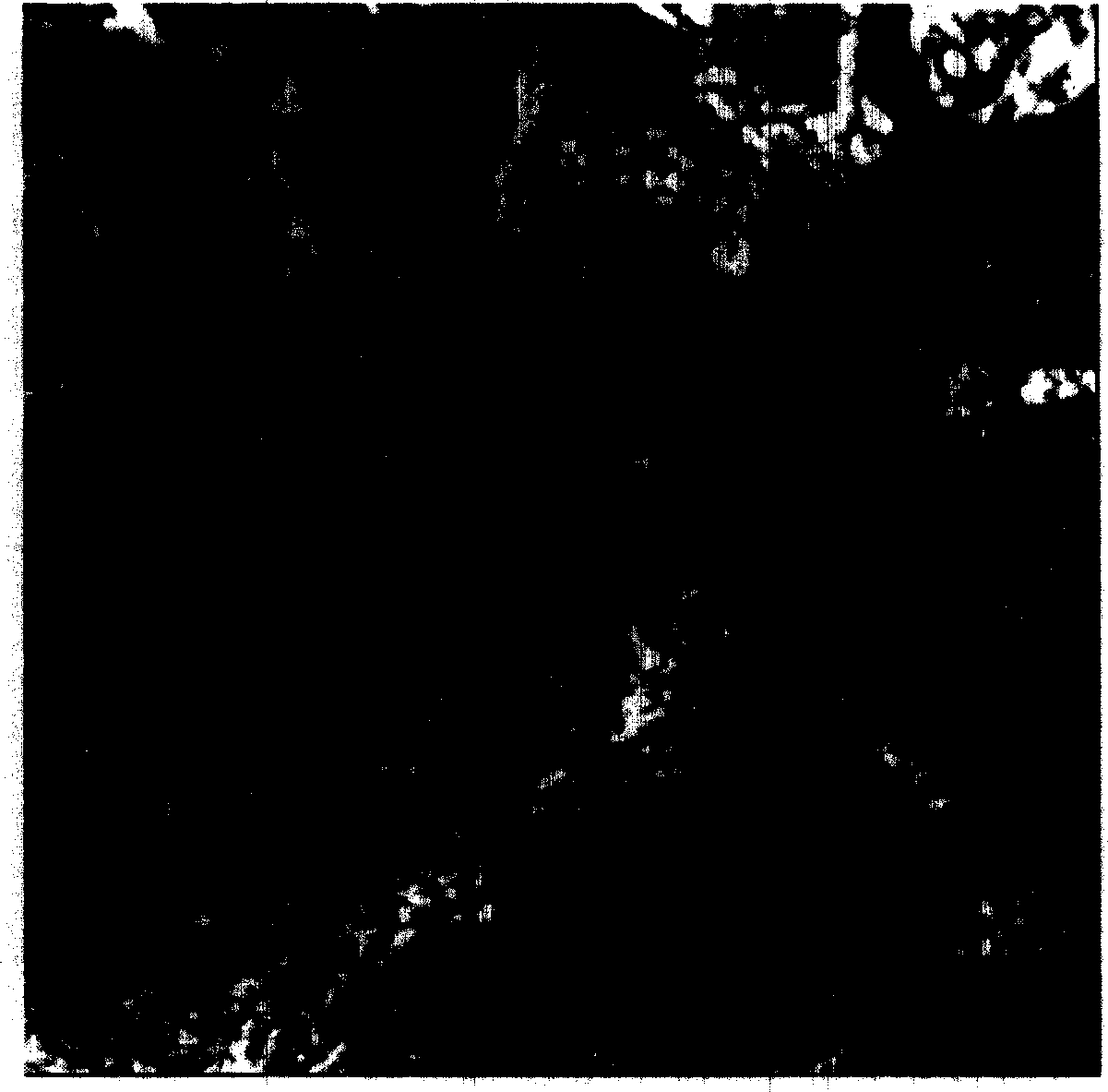
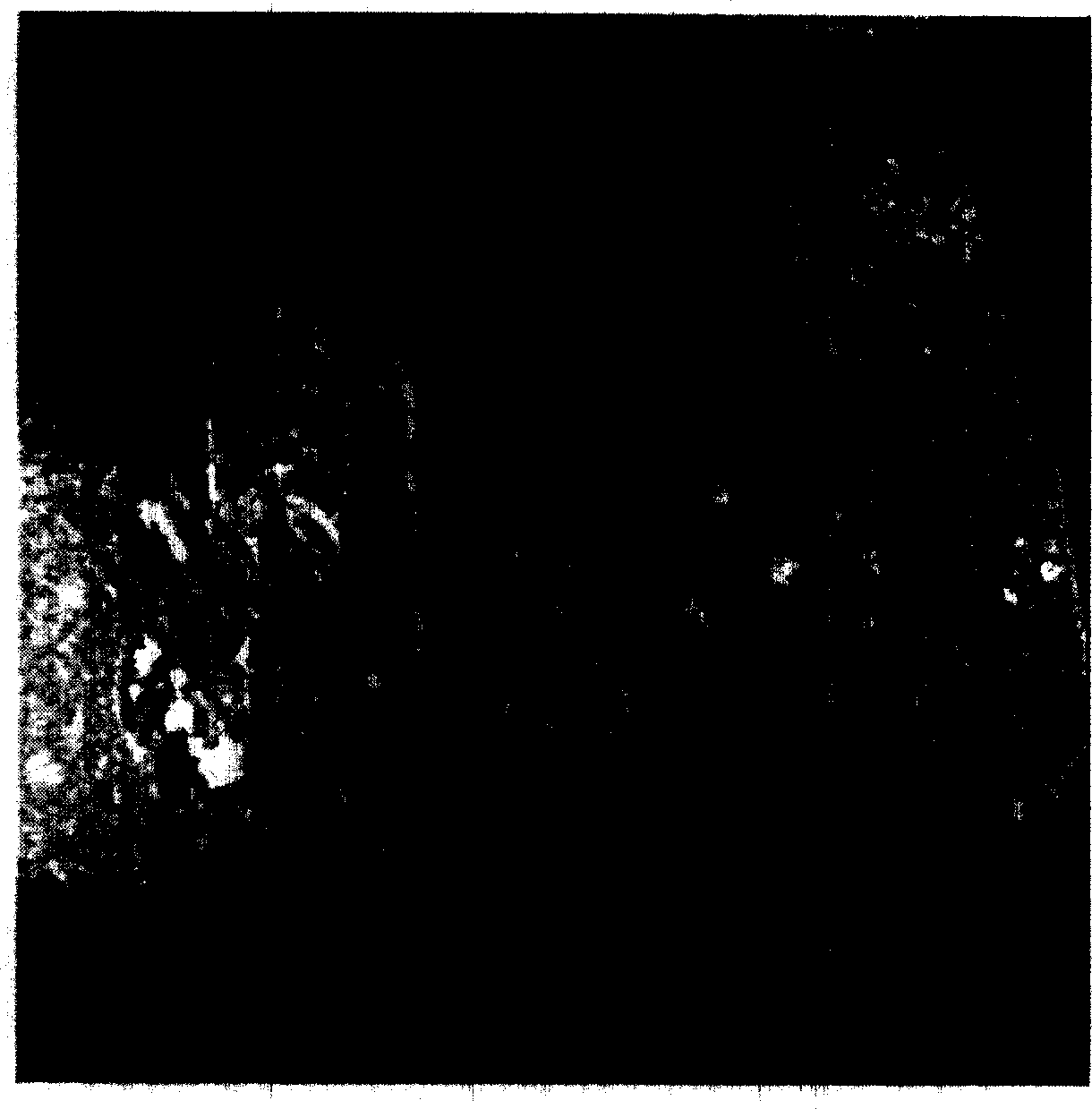
[0018] Human tongue squamous cell carcinoma tissue samples were taken from the clinical operation of oral and maxillofacial surgery in a hospital, cut into small pieces less than 2 mm square, and fixed with 2% glutaraldehyde fixative solution pre-cooled in ice water for 2 hours. The fixative formula is 50ml of 0.2mol / L phosphate buffer solution, 8ml of 25% glutaraldehyde aqueous solution, add ultrapure water to 100ml, and put it in an ice-water bath to cool it for later use. Then wash twice with 0.2mol / L phosphate buffer, and then carry out ethanol gradient dehydration. The dehydration process is as follows:
[0019] Under freezing conditions (0°C-4°C), 50% ethanol water solution for 15 minutes, 70% ethanol water solution for 15 minutes, and 95% ethanol water solution for 15 minutes at room temperature, anhydrous ethanol was replaced once, each time 10 minutes Minutes, absolute ethanol was replaced once, every 10 minutes.
[0020] After the sample has been dehydrated, it is d...
Embodiment 2
[0023]After culturing C6 cells, pour out the culture medium, first wash twice with 0.2mol / L phosphate buffer solution, then add ice water pre-cooled 2% glutaraldehyde fixative solution to fix at low temperature for 1 hour, and centrifuge the obtained cell mass with ethanol The solution is subjected to gradient dehydration, followed by 50%→70%→95%→absolute ethanol→absolute ethanol, 10 minutes each time, in which absolute ethanol and absolute ethanol should be replaced once respectively, and ice water is required for 50% and 70% ethanol Pre-chill and dehydrate at low temperature. After the sample was dehydrated, it was directly transferred to the liquid with a volume ratio of epoxy resin 618: absolute ethanol = 1:1 and placed on a shaker to infiltrate for 1 hour, then poured out the liquid, blotted the residue with filter paper, and then added ring Oxygen resin 618 mixed solution, infiltrated in a 35°C incubator for 3 hours. Shake a few times in the middle to facilitate penetra...
Embodiment 3
[0026] After Tca8113 cells are cultured, discard the culture medium, rinse with 0.2mol / L phosphate buffer, add ice water pre-cooled 2% glutaraldehyde fixative solution to fix at low temperature for 1.5 hours, and dehydrate the cell mass obtained after centrifugation with gradient ethanol solution , followed by 50%→70%→95%→absolute ethanol→absolute ethanol, every 12 minutes, anhydrous and absolute ethanol are replaced once, 50% and 70% ethanol need to be pre-cooled with ice water and carried out at low temperature dehydration. After the sample is dehydrated, directly use the epoxy resin 618 mixed solution: absolute ethanol = 1:1 volume ratio liquid to infiltrate on the shaker for 1.5 hours, then pour the solution, absorb the residue with filter paper, and then add epoxy Resin 618 mixed solution, infiltrated in an incubator at 35°C for 3.5 hours. Pick out the sample, carefully absorb the residue with filter paper, and embed it with epoxy resin 618 mixture. Then heat polymeriza...
PUM
 Abstract
Description
Claims
Application Information
 - R&D
- Intellectual Property
- Life Sciences
- Materials
- Tech Scout
- Unparalleled Data Quality
- Higher Quality Content
- 60% Fewer Hallucinations
Browse by: Latest US Patents, China's latest patents, Technical Efficacy Thesaurus, Application Domain, Technology Topic, Popular Technical Reports.
© 2025 PatSnap. All rights reserved.Legal|Privacy policy|Modern Slavery Act Transparency Statement|Sitemap|About US| Contact US: help@patsnap.com


