Organism tissue staining method

A dyeing method and biological technology, applied in biochemical equipment and methods, biological testing, microbial measurement/inspection, etc., can solve the problems of unsuitable living tissue dyeing, toxicity of dyeing reagents, cumbersome steps, etc., and achieve bright colors and outlines. Clear, time-saving results

Inactive Publication Date: 2003-09-10
PEKING UNIV
View PDF0 Cites 4 Cited by
  • Summary
  • Abstract
  • Description
  • Claims
  • Application Information

AI Technical Summary

Problems solved by technology

However, the disadvantages of traditional staining methods are that the steps are cumbersome and time-consuming, and the samples often need special treatment; in addition, the staining reagents are highly toxic and generally not suitable for living tissue staining

Method used


Image

  • Organism tissue staining method
  • Organism tissue staining method
  • Organism tissue staining method

Examples


Embodiment 1

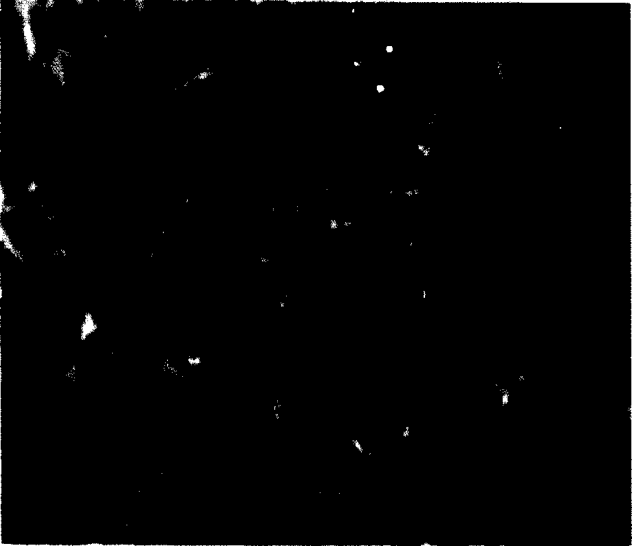
[0041] Prepare the fluorescent complex europium-thienoyltrifluoroacetone-triphenylphosphine oxide into a 5 mg / L ethanol solution, drop it on the phospholipid, wash it with distilled water after one minute, and then observe it with a fluorescent microscope , the phospholipids are colored into purple clumps, such as figure 1 shown.

Embodiment 2

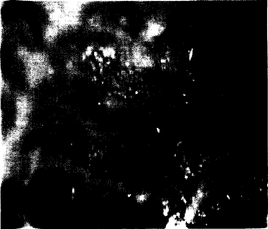
[0043] Using a method similar to Example 1, the mucin was stained with a terbium-ciprofloxacin solution with a concentration of 1 mg / liter for 3 minutes. Under the excitation of ultraviolet light, bright green fluorescence appeared, such as figure 2 shown.

Embodiment 3

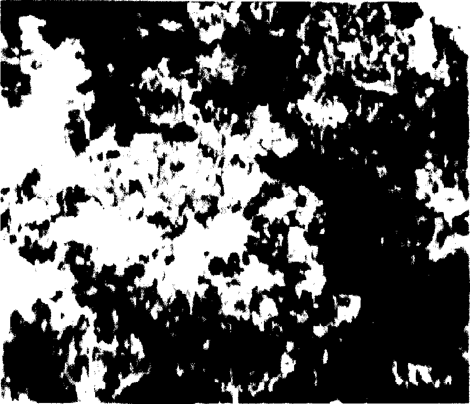
[0045] With a method similar to Example 1, the terbium-ofloxacin solution with a concentration of 10 mg / liter makes sodium nucleotide staining, staining for 3 minutes, under the excitation of ultraviolet light, bright red fluorescent patterns appear, such as image 3 shown.


PUM

PropertyMeasurementUnit
concentrationaaaaaaaaaa

Abstract

The present invention relates to organism tissue staining method with RE fluorescent complex. The staining reagent used in the present invention is solution of RE fluorescent complex, and the solution is dropped onto the organism tissue or the test sample is soaked in the solution for 0.5-3 min before flushing with distilled water. Under the exciting of ultraviolet light, the configuration and detailed structure of the stainless tissue sample may be observed in fluorescent microscope. The said method has simple operatino and short period, and the sample needs no pre-treatment and unlimited. The reagent has no or only weak toxicity and the staining process has no influence on the structure of tissue or cell. The tissue may be extracorporeal or intracorporeal.

Description

Technical field: [0001] The invention relates to a biological tissue dyeing method, in particular to a method for using a rare earth fluorescent complex as a dye to dye biological tissue. Background technique: [0002] Since the discovery of the cells that make up the human body, the understanding of the microstructure of organisms represented by the human body has always been an important research field. The use of chemical substances to stain cells or tissues is an important research method, from histochemical staining represented by Hematoxylin and Eosin staining (HE) to immunohistochemical staining of monoclonal antibody labeling. Staining greatly improves the resolution ability, and at the same time, broadens the observation level. With the development of fluorescent reagents and fluorescent observers, fluorescent histochemistry and immunofluorescence staining are widely used in life science research with their unique colors. field and become a hot research topic. How...

Claims


Application Information

Patent Timeline
Patent Type & Authority Applications(China)
IPC IPC(8): C12Q1/00G01N1/30G01N33/48
Inventor 吴瑾光徐怡庄翁诗甫吴奇光王晶杨军杨丽敏周风山杨展澜李维红苏允兰王凡张莉任宇刘智
Owner PEKING UNIV
Why Eureka
Try Eureka