Low virulent strain of recombinant newcastle disease lasota vaccine expressing HA protein of avian influenza-H5 virus
A technology of avian influenza virus and attenuated vaccine, which is applied to a method and the recombinant Newcastle disease LaSota attenuated vaccine strain in the field of preparation and prevention of poultry, HA and rLa, which can solve the problems of inconvenient use, high manufacturing cost, and unrealized wide application.
- Summary
- Abstract
- Description
- Claims
- Application Information
AI Technical Summary
Problems solved by technology
Method used
Image
Examples
Embodiment 1
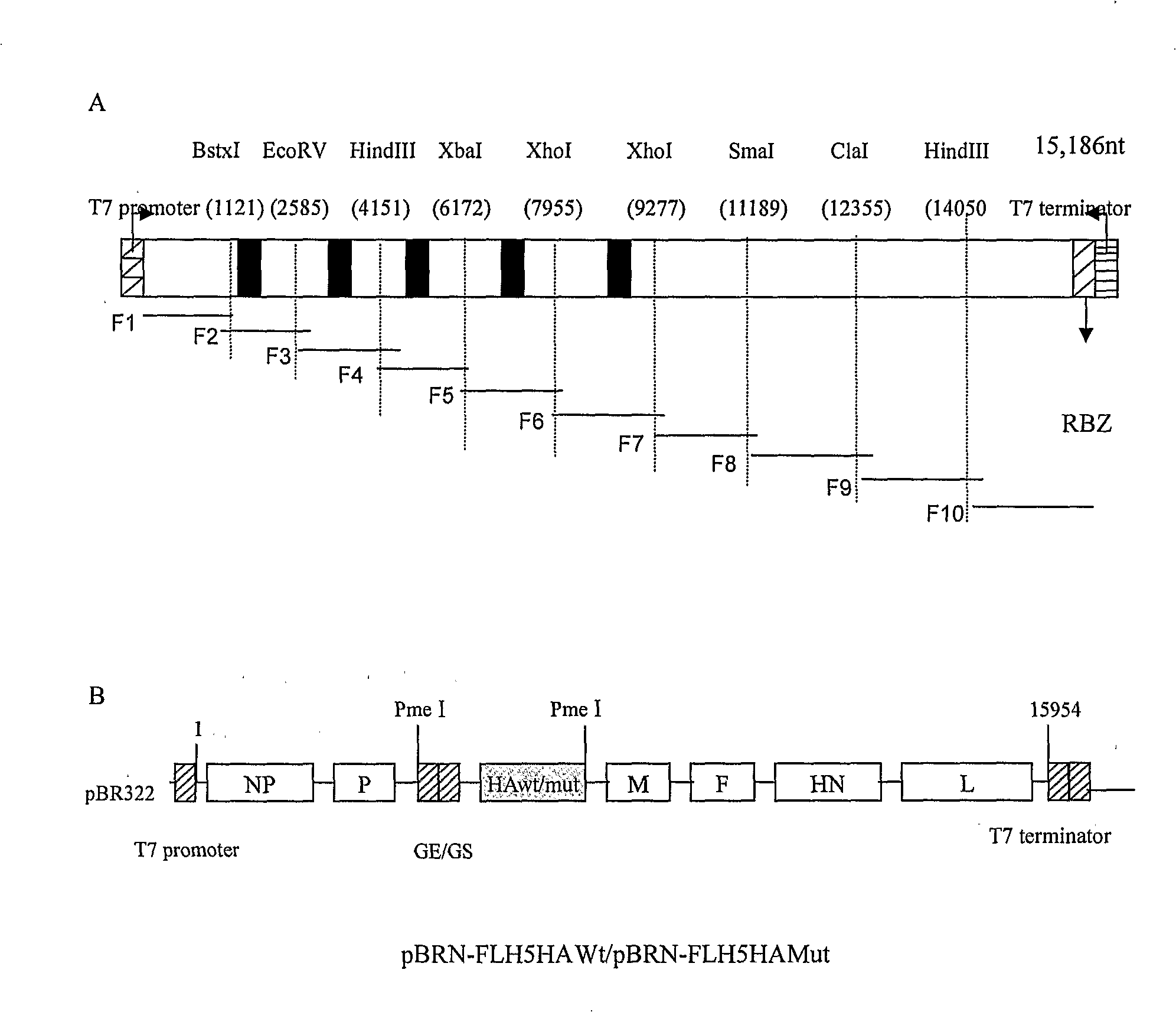
[0036] Example 1 Construction of recombinant Newcastle disease LaSota attenuated vaccine strain expressing wild-type or mutant avian influenza virus H5 subtype hemagglutinin (HA) protein
[0037] Cells, viruses and test materials
[0038] BHK-21 cells (milk hamster kidney cells ATCC CCL-10), culture medium is DMEM (Dulbecco's modified Eagle's medium) containing 10% fetal bovine serum (Hyclone) and 1 μg / ml G418; NDV Lasota vaccine Strain AV1615 (purchased from China Center for Veterinary Culture Collection (CVCC)). The allantoic cavity of 9-10-day-old SPF chicken embryos was inoculated and frozen at -70°C for later use; chicken anti-NDV hyperimmune serum was prepared by our laboratory (Chu, H.P., G.Snell, D.J.Alexander, and G.C.Schild.1982 .Avian Pathol 11:227-234); SPF chicken embryos and SPF chicks were provided by the SPF Experimental Animal Center of Harbin Veterinary Research Institute. H5 subtype highly pathogenic avian influenza virus (HPAIV) A / Goose / Guangdong / 96 / 1 / H5N...
Embodiment 2
[0047] Embodiment 2 Recombinant NDV expresses AVI HA protein indirect immunofluorescence assay (IFA) test
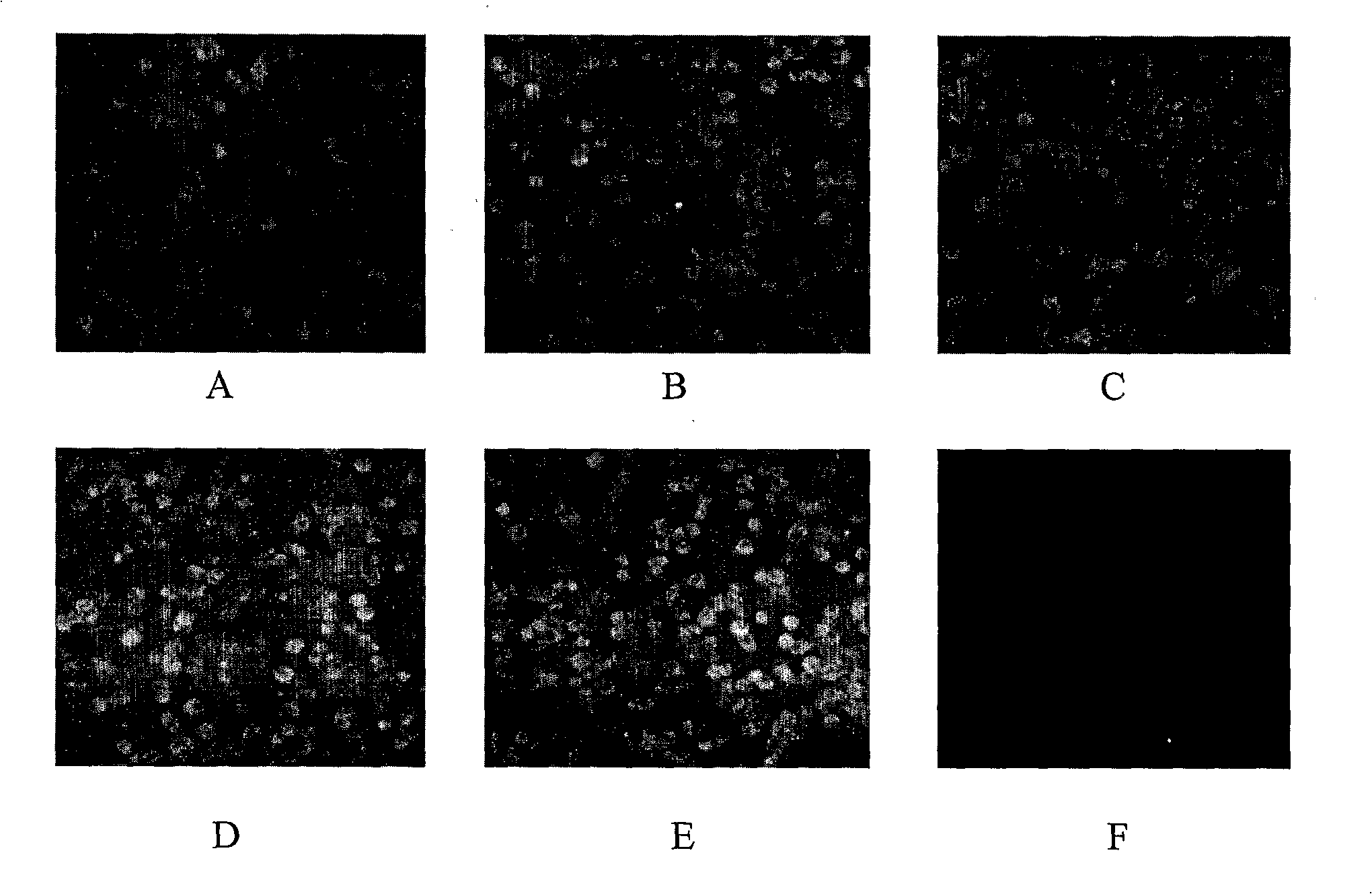
[0048] The NDV LaSota vaccine strain can transiently infect mammalian cells cultured in vitro. In order to prove the replication of rLasota-H5wtHA and rLasota-H5mutHA viruses in BHK-21 cells and the expression of virus antigens, the two allantoxins infected about 70-80% of monolayer BHK-21 cells with MOI as 1 virus load ( image 3 A and B), while using the NDV wild-type LaSota vaccine strain infected cells as a control ( image 3 C), 20 hours after the infection, the early CPE (cytopathic) phenomenon appeared in the cells, and immediately carried out indirect immunofluorescence staining with the positive serum of NDV high-immune SPF chickens as the detection antibody. As a result, strong positive reactions were observed under the fluorescence microscope of three kinds of virus-infected cells ( image 3 A, B and C) More specifically, the experimental steps are as follows...
Embodiment 3
[0051] Example 3 Western-Blot identification of recombinant NDV expressing H5 subtype avian influenza virus HA protein
[0052] Take virus-infected chicken embryo fibroblast (CEF) lysate (after discarding the culture medium, add 1 / 10 volume of PBS, suspend the cells, add an equal volume of 2×SDS lysis buffer to lyse in boiling water for 10 min, centrifuge at 12000 g for 10 min , harvest the supernatant) or virus-inoculated SPF chicken embryo allantoic stock solution, carry out SDS-PAGE (Bio-Rad). Protein electrotransfer (Bio-Rad) to nylon membrane (Ameresco), 10% skimmed milk blocked overnight, PBST (0.05% Tween20) was added after washing, 1:50 diluted DNA was added for immunization to prepare chicken anti-H5 subtype avian influenza virus HA Antigen hyperimmune serum was the primary antibody, horseradish peroxidase (HRP)-labeled rabbit anti-chicken goat anti-mouse IgG (Sigma) was the secondary antibody, diluted 1:2500 times in PBST, DAB (diaminobenzidine, Sigma) After 3-5 min...
PUM
 Abstract
Description
Claims
Application Information
 - R&D
- Intellectual Property
- Life Sciences
- Materials
- Tech Scout
- Unparalleled Data Quality
- Higher Quality Content
- 60% Fewer Hallucinations
Browse by: Latest US Patents, China's latest patents, Technical Efficacy Thesaurus, Application Domain, Technology Topic, Popular Technical Reports.
© 2025 PatSnap. All rights reserved.Legal|Privacy policy|Modern Slavery Act Transparency Statement|Sitemap|About US| Contact US: help@patsnap.com


