
Ultrafine nanoparticles as multimodal contrast agent

a technology of nanoparticles and contrast agents, applied in the direction of capsule delivery, microcapsules, drug compositions, etc., can solve the problems of low survival rate of patients suffering from lung cancer, inability to optimally use negative contrast agents for imaging the lung, and inability to meet the needs of patients with lung cancer

Active Publication Date: 2019-12-31
UNIV DE BORDEAUX +4
View PDF8 Cites 0 Cited by
  • Summary
  • Abstract
  • Description
  • Claims
  • Application Information

AI Technical Summary

Benefits of technology

[0023](iv) to propose novel means of administering a prodrug or a contrast agent, requiring few devices and making it possible to reduce the doses to be injected by a route that is noninvasive, comfortable and nontoxic for the patient;
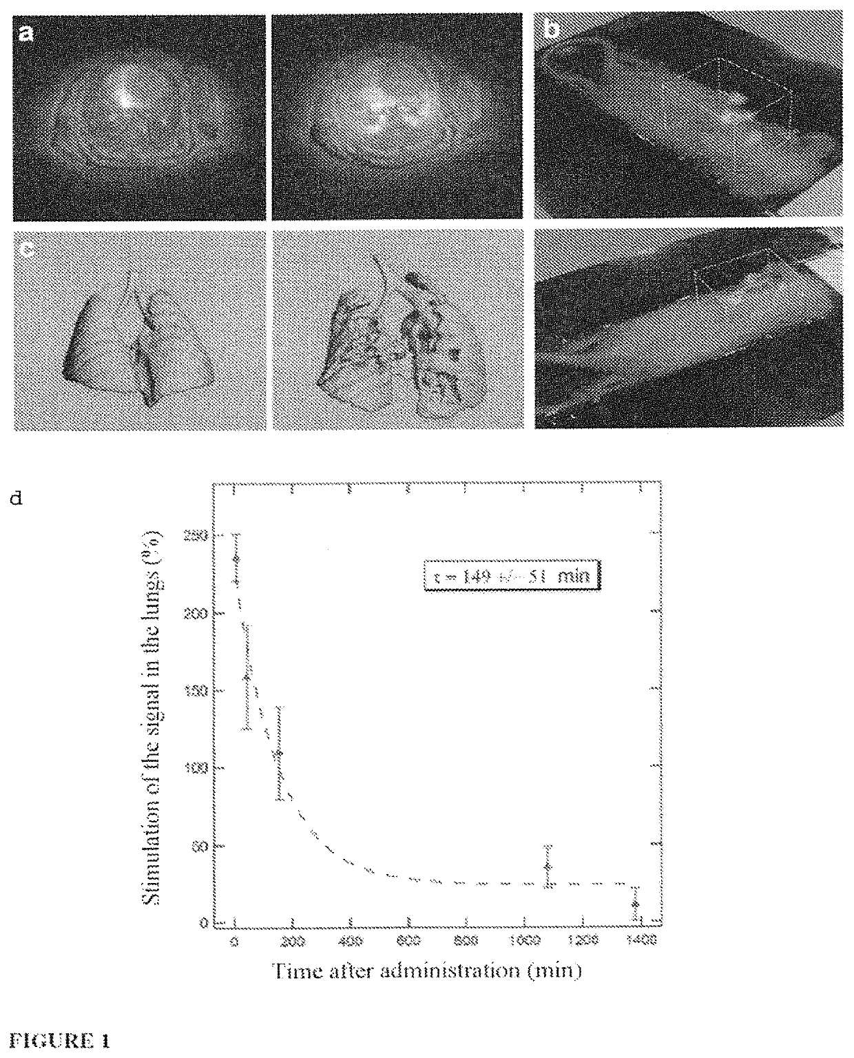
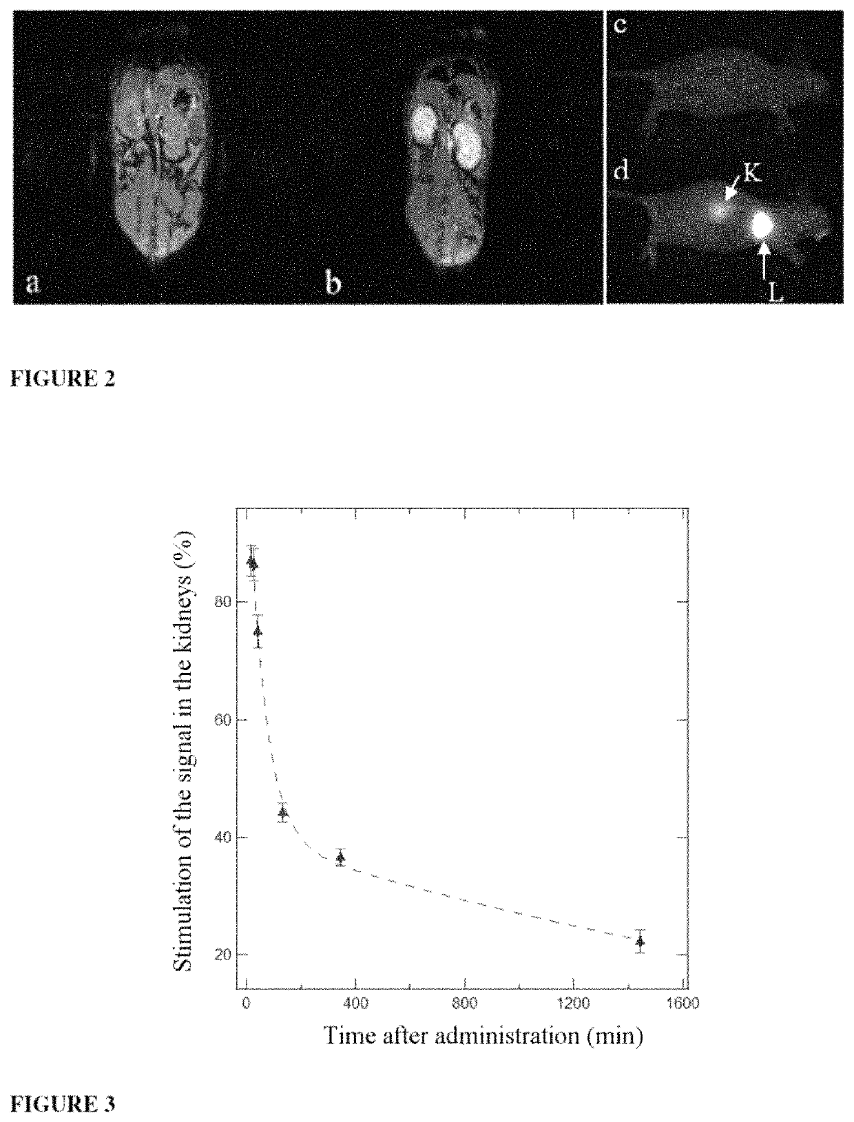
[0180]The mode of administration via the airways enables a local administration in the lungs, but also passage of the nanoparticles in the systemic circulation. Thus, by coupling therapeutic molecules onto the nanoparticles (covalent or noncovalent coupling), the invention proposes the use of these nanoparticles as a vector for the administration via the airways, in the blood stream, of therapeutic molecules or of contrast agents. According to the mode of coupling of the therapeutic molecules, these vectors can be used as a prodrug with controlled, sustained or delayed release of the active ingredient.

Problems solved by technology

The low survival of patients suffering from lung cancer is mainly due to the absence of tools for early diagnosis and for locally targeting therapeutic agents.
However, the administration of nanoparticles that can be used as a contrast agent is conventionally carried out by intravenous injection owing to the risks of long-term retention in the lungs (and therefore of associated tonicity) (J. Roller et al., Nanomedicine, 2011, 7, 753-762 / R.
However, the use of a negative contrast agent is not optimal for imaging the lung, and the diagnosis might be improved by using positive contrast agents.
For fluorescence imaging, near-infrared (NIR) emission nanoparticles, such as quantum dots, have been used, but their inherent toxicity limits the possible future clinical applications. A. Zintcheko et al., Molecular Therapy, 2009, 17, 1849-1856 / F-X.
This approach does not make it possible to obtain a strong MRI signal (4 Gd3+ for molecules of more than 10 kDa) or else it is necessary to dextran polymers of much higher molar mass (greater than 70 kDa).
The contrast agent is eliminated by expectoration and does not make it possible to obtain an enhancement of the pulmonary tissues.

Method used


Image

  • Ultrafine nanoparticles as multimodal contrast agent
  • Ultrafine nanoparticles as multimodal contrast agent
  • Ultrafine nanoparticles as multimodal contrast agent

Examples


example 1

Synthesis of Hybrid Gadolinium Nanoparticles of Gd—Si-DOTA: SRP (Small Rigid Platforms) Type

[0200]The nanoparticles are obtained by means of a three-stage synthesis as described in WO 2011 / 135101. First of all, the oxide cores are synthesized in diethylene glycol before growth of the polysiloxane layer, followed by the covalent grafting of completing agents to the surface of the nanoparticle; the gadolinium oxide core dissolves when placed in water, thereby leading to fragmentation of the particles which makes it possible to obtain small particles of less than 5 nanometers composed only of polysiloxane, or organic species and of complexing agents useful in imaging.

[0201]The advantage of this top down approach is to make it possible to obtain very small particles which have multimodal characteristics while at the same time having the advantage of being easy to eliminate via the kidneys.

Detailed Description of the Synthesis

Oxide Cores

[0202]A first solution is prepared by dissolving 5....

example 2

Functionalization of the Nanoparticles for Active Targeting of Tumors: SRP-cRGD

[0206]The nanoparticles used for the grafting are similar to those described in example 1. They therefore possess a set of free DOTAs (and thus of available carboxylic acid functions) for the grafting of cRGD which is known to be an agent that targets αvβ3 integrin. The grafting of cRGD to the particle is carried out by means of peptide coupling between a carboxylic acid function of a DOTA unit and the primary amine function of the cRGD peptide. The nanoparticles obtained in example 1 are diluted in water (Gd3+ concentration of around 100 mM). A mixture of EDC and NHS (3.4 EDC and 3.4 NHS per Gd) is added to the particles, the pH is adjusted to 5 and the mixture is kept stirring for 30 minutes. The cRGD (2.3 cRGD per Gd) is dissolved separately in anhydrous DMSO. It is then added to the previous solution and the pH of the mixture is adjusted to 7.1 before leaving it to stir for 8 hours. The solution is fi...

example 3

Analysis of the Interaction of SRPcRGD Nanoparticles Obtained According to Example 2 with Cells Expressing αvβ3 Integrin by Flow Cytometry and Fluorescence Microscopy

Analysis by Fluorescence Microscopy

[0207]HEK293(β3) cells, which overexpress αvβ3 integrin, are cultured on coverslips in 12-well plates (100 000 cells per well) overnight at 37° C. They are rinsed once with 1×PBS, then with 1×PBS containing 1 mM of CaCl2 and 1 mM of MgCl2. They are then incubated for 30 minutes at 4° C. (binding analysis) or 30 minutes at 37° C. (internalization analysis), in the presence of SRP (nanoparticles according to example 1) with a fluorophore of Cy5.5 type, of SRP-cRGD (obtained according to example 2) Cy5.5 or of SRP-cRAD (obtained according to example 2 and acting as a negative control) Cy5.5 at a Gd3+ ion concentration of 0.1 mM. They are then rinsed with PBS Ca2+ / Mg2+ (1 mM) and fixed (10 minutes with 0.5% paraformaldehyde. The nuclei are labeled with Hoechst 33342 (5 μM for 10 minutes) (...


PUM

PropertyMeasurementUnit
mean diameteraaaaaaaaaa
diameteraaaaaaaaaa

Abstract

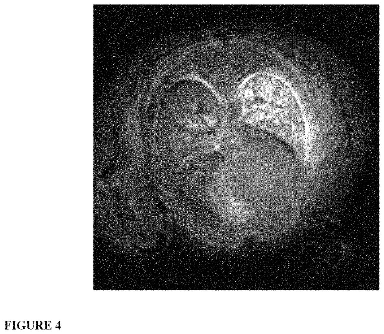
The invention relates to a novel use of ultrafine nanoparticles, of use as a diagnostic, therapeutic or theranostic agent, characterized by their mode of administration via the airways. The invention is also directed toward the applications which follow from this novel mode of administration, in particular for imaging the lungs, and the diagnosis or prognosis of pathological pulmonary conditions. In the therapeutic field, the applications envisioned are those of radiosensitizing or radioactive agents for radiotherapy (and optionally curietherapy), or for neutron therapy, or of agents for PDT (photodynamic therapy), in particular for the treatment of lung tumors.

Description

TECHNICAL FIELD[0001]The invention relates to a novel use of ultrafine nanoparticles, as a diagnostic, therapeutic or theranostic agent, characterized by their mode of administration via the airways. The invention is also directed toward the applications which follow from this novel mode of administration, in particular for imaging the lungs, and the diagnosis or prognosis of pathological pulmonary conditions. In the therapeutic field, the applications envisioned are those of radiosensitizing or radioactive agents for radiotherapy (and optionally curietherapy), or for neutron therapy, or of agents for phototherapy or thermotherapy, in particular for the treatment of lung tumors.TECHNOLOGICAL BACKGROUND[0002]Despite considerable research efforts, cancer remains one of the major causes of death throughout the world (R. Siegel et al.: Cancer Statistics 2012, 2012, 62, 10-29). Among the various types of cancer, lung cancer constitutes the principal cause of cancer-associated death (J. S...

Claims


Application Information

Patent Timeline
Patent Type & Authority Patents(United States)
IPC IPC(8): A61K47/69A61K49/18A61K49/10A61K49/00A61K49/04A61K41/00A61K51/12A61K9/51
CPCA61K49/183A61K47/6929A61K49/108A61K41/0038A61K41/0057A61K49/0017A61K49/0032A61K47/6923A61K49/105A61K51/1244A61K9/5146A61K49/04A61K49/00A61K49/1824A61P11/06A61P35/00
Inventor CREMILLIEUX, YANNICKBIANCHI, ANDREADUFORT, SANDRINECOLL, JEAN-LUCLUX, FRANCOISTILLEMENT, OLIVIER
Owner UNIV DE BORDEAUX
Learn More