
Method for culture of stem cell

a stem cell and culture method technology, applied in the field of stem cell culture, can solve the problems of difficult control of selective differentiation from those progenitor cells to particular cerebral cortical neurons, difficult to obtain human cerebral tissue for the sake of these researches, and inability to efficiently induce in vitro differentiation. , to achieve the effect of efficiently induced stem cell differentiation, selectively induced differentiation of layer-specific neurons, and efficient induction of differentiation

Active Publication Date: 2021-03-02
RIKEN CO LTD
View PDF29 Cites 1 Cited by
  • Summary
  • Abstract
  • Description
  • Claims
  • Application Information

AI Technical Summary

Benefits of technology

This approach enables high-efficiency induction of cerebral cortex progenitor cells and specific neuronal types, such as hypothalamic neurons, with improved control over differentiation pathways, enhancing the potential for regenerative medicine and drug discovery applications.

Problems solved by technology

To this end, disease-related nerve cells that are present in the brain must be produced accurately; however, because an extremely large number of kinds of nerve cells are present in the brain, there are still many types of nerve cells and brain tissues for which efficient in vitro differentiation has been unsuccessful.
For the treatment of cerebral disorders, etiologic research, drug discovery research, cell transplantation therapy research and the like have been conducted so far, but it is extremely difficult to obtain human cerebral tissue for the sake of these researches.
Although it has recently become possible to induce differentiation of an embryonic stem cell into cortical progenitor cells (see Non-Patent Document 4), it has been difficult to control the selective induction of differentiation from those progenitor cells to particular cerebral cortical neurons.
Furthermore, the majority of the cerebral tissues induced by a conventional method such as the SFEB method failed to have a clear morphology of cortical tissue, mostly forming disarrayed cell masses.
Additionally, the conventional SFEB method does not enable efficient induction of differentiation of the tissue of the rostral diencephalon, which develops on the most rostral side of the central nervous system, particularly of the hypothalamus, from an ES cell.

Method used


Image

  • Method for culture of stem cell
  • Method for culture of stem cell
  • Method for culture of stem cell

Examples


example 1

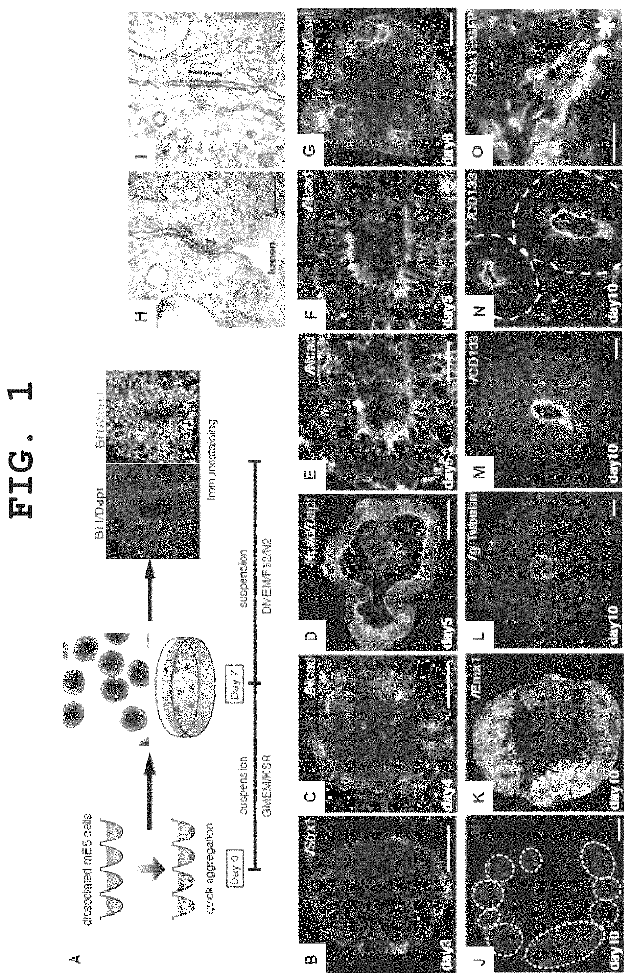
Highly Efficient Differentiation Induction into Cerebral Cortex Progenitor Cells by the SFEBq Method

(Method)

[0258]EB5 cells, which are mouse ES cells (E14-derived), or cells of an E14-derived cell line wherein the Venus gene, which is a modified GFP (green fluorescent protein), has been knocked in the cerebral nerve marker Bf1 gene as a nerve differentiation reporter by homologous recombination (hereinafter described as “Bf1 / Venus-mES cells”), were cultured as described in the literature (Watanabe et al., Nature Neuroscience, 2005), and used in the experiments.

[0259]The medium used was a G-MEM medium (Invitrogen) supplemented with 1% fetal calf serum, 10% KSR (Knockout Serum Replacement; Invitrogen), 2 mM glutamine, 0.1 mM non-essential amino acids, 1 mM pyruvic acid, 0.1 mM 2-mercaptoethanol and 2000 U / ml LIF. For nerve differentiation induction by suspension culture, ES cells were mono-dispersed using 0.25% trypsin-EDTA (Invitrogen), and suspended in 150 μl of the differentiation ...

example 2

In Vitro Production of Cerebral Neurons from Cerebral Cortex Progenitor Cells Induced by the SFEBq Method

(Method)

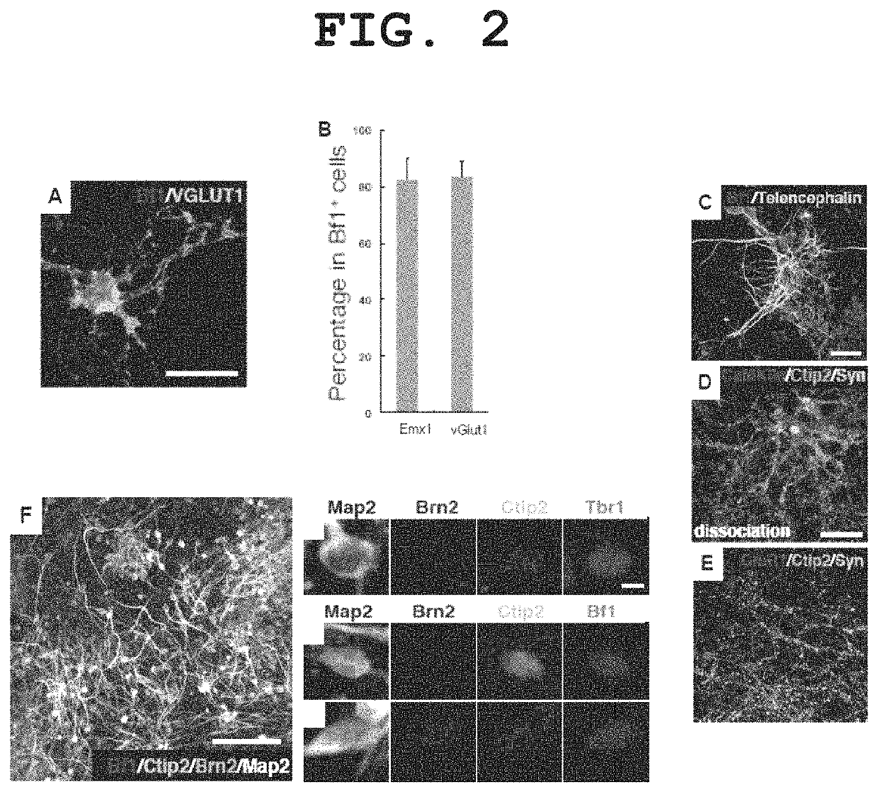
[0264]Aggregates obtained by continued differentiation culture by the method described in Example 1 for 12 days were enzymatically dispersed (SUMILON Neural Tissue Dissociation kit), plated in a culture plate coated with poly-D-lysine / laminin / fibronectin at 5×10⁴ cells / cm2, and cultured using a DMEM / F12 medium supplemented with 1×N2 supplement and 10 ng / ml of FGF2 for 2 days. Subsequently, the cells were further cultured using a Neurobasal medium supplemented with B27 supplement+50 ng / ml BDNF+50 ng / ml NT3 for 6 days. The properties of the differentiated neurons were analyzed by a fluorescent immunostaining method. The results are shown in FIG. 2.

(Results)

[0265]Most of the cells in the test tube became TuJ1-positive neurons, of which 80% were positive for the cerebral cortex-specific marker Emx1 and positive for the glutamatergic neuron (abundantly present in cerebral cort...

example 3

Formation of Nerve Network by Cerebral Neurons Produced by the SFEBq Method

(Method)

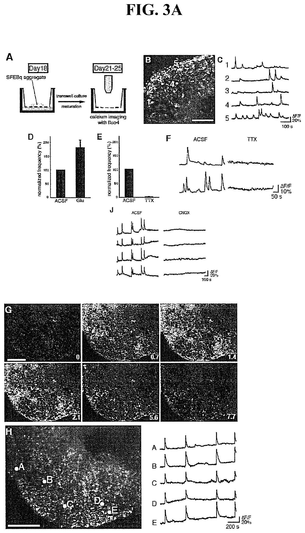
[0267]To confirm the activities and network formation of cerebral neurons produced by the SFEBq method, an analysis was performed by a Ca++ imaging method using fluo4-AM. The method of examination was as described in the literature (Ikegaya et al, 2005, Neurosci. Res., 52, 132-138).

[0268]Aggregate masses derived from a mouse ES cell cultured by the method described in Example 1 for 18 days were further cultured on the Transwell culture insert (Corning) using a DMEM / F12 medium supplemented with N2 for 7 days (FIG. 3A, view A). Ca++ imaging was performed at room temperature using an artificial cerebrospinal fluid. The results are shown in FIGS. 3A and 3B.

(Results)

[0269]In the Ca++ imaging, many cells exhibited repeatedly observable Ca++ elevations synchronized or not synchronized with surrounding cells (FIG. 3A, views B and C). Since all these elevations were enhanced by administration of glutamine (FIG...


PUM


Abstract

The present invention enables efficient suspension culture of stem cells in a serum-free medium by comprising a step for quickly forming a homogenous aggregate of stem cells, and provides a method of selectively inducing the differentiation of nerves from a stem cell, a method of forming a cerebral cortical nerve network in vitro, and a method of producing a steric structure of a brain tissue in vitro, as well as a method of producing hypothalamic neuron progenitor cells, comprising culturing pluripotent stem cells as a suspended aggregate in a serum-free medium that substantially does not contain a Nodal signal promoter, a Wnt signal promoter, an FGF signal promoter, a BMP signal promoter, retinoic acid and an insulin, and isolating hypothalamic neuron progenitor cells from the culture.

Description

CROSS-REFERENCE TO RELATED APPLICATIONS[0001]This patent application is a continuation of co-pending U.S. patent application Ser. No. 12 / 996,503, filed on Dec. 6, 2010, which is the U.S. national phase of International Patent Application No. PCT / JP2009 / 060396, filed Jun. 5, 2009, which claims the benefit of Japanese Patent Application No. 2008-149880, filed on Jun. 6, 2008, and Japanese Patent Application No. 2008-282299, filed on Oct. 31, 2008, which are incorporated by reference in their entireties herein.TECHNICAL FIELD[0002]The present invention relates to a method of culturing stem cells. Specifically, the present invention relates to a method of inducing differentiation of stem cells comprising combining quick re-aggregation and three-dimensional suspension culture in performing stem cell aggregate culture, and to a cell culture obtained by the method, and the like.BACKGROUND ART[0003]To date, some culturing methods for inducing differentiation of nerves from a pluripotent ste...

Claims


Application Information

Patent Timeline
Patent Type & Authority Patents(United States)
IPC IPC(8): C12N5/0797
CPCC12N5/0623C12N2501/33C12N2501/415C12N2501/42C12N2501/70C12N2506/02
Inventor SASAI, YOSHIKIWATAYA, TAKAFUMIEIRAKU, MOTOTSUGU
Owner RIKEN CO LTD
Learn More